Researchers from Vanderbilt University report that when people having their brains scanned by fMRI perform a task, such as wiggling their fingers, blood oxygenation-level dependent (BOLD) signals increase in white matter throughout the brain.
“We don’t know what this means. We just know that something is happening. There truly is a powerful signal in the white matter,”
said first author, Kurt Schilling, Ph.D., research assistant professor of Radiology and Radiological Sciences at Vanderbilt.
White Matter Activity
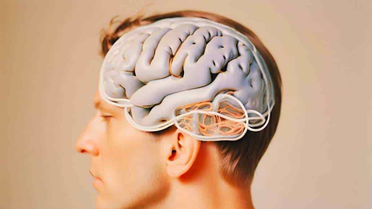
The human brain is composed of two types of matter: nerve cell bodies (gray matter) that process sensation, control voluntary movement, and enable speech, learning, and cognition, and axons (white matter) that connect cells and project to the rest of the body.
Historically, scientists have concentrated on the gray matter of the cortex, figuring that’s where the action is, while ignoring white matter, even though it makes up half the brain. Researchers at the Vanderbilt University Institute of Imaging Science, University are out to change that.
Schilling stated that it is crucial to pursue these latest findings because a variety of neurological disorders, including epilepsy and multiple sclerosis, disrupt the “connectivity” of the brain. This suggests that white matter activity is occurring.
The researchers will continue to investigate the alterations in white matter signals that they have previously identified in schizophrenia, Alzheimer’s disease, and other brain diseases. In addition, they aim to identify the biological basis for these changes through animal studies and tissue analysis.
Increased Blood Flow
BOLD signals in gray matter indicate an increase in blood flow (and oxygen) in response to increased nerve cell activity.
Perhaps axons or the glial cells that maintain their protective myelin sheath use more oxygen when the brain is “working” as well. Or perhaps these signals are in some way related to gray matter activity.
Even if nothing biological is happening in white matter, Schilling believes something is going on.
“The signal is changing. It’s changing differently in different white matter pathways and it’s in all white matter pathways, which is a unique finding,”
he said.
Signal to Noise
One reason that white matter signals have been understudied is that they have lower energy than gray matter signals, and thus are more difficult to distinguish from the brain’s background “noise.”
The researchers increased the signal-to-noise ratio by having the person whose brain was being scanned repeat a visual, verbal, or motor task many times in order to establish a trend and by averaging the signal across many different white matter fiber pathways.
“For 25 or 30 years, we’ve neglected the other half of the brain,”
Schilling said. White matter signals have not only been ignored by some researchers, but they have also been removed from their reports on brain function.
The researchers concluded that the Vanderbilt findings suggest that many fMRI studies may not only underestimate the true extent of brain activation, but may also omit vital information from the MRI signal.
Abstract
Recent studies have revealed the production of time-locked blood oxygenation level–dependent (BOLD) functional MRI (fMRI) signals throughout the entire brain in response to tasks, challenging the existence of sparse and localized brain functions and highlighting the pervasiveness of potential false negative fMRI findings. “Whole-brain” actually refers to gray matter, the only tissue traditionally studied with fMRI. However, several reports have demonstrated reliable detection of BOLD signals in white matter, which have previously been largely ignored. Using simple tasks and analyses, we demonstrate BOLD signal changes across the whole brain, in both white and gray matters, in similar manner to previous reports of whole brain studies. We investigated whether white matter displays time-locked BOLD signals across multiple structural pathways in response to a stimulus in a similar manner to the cortex. We find that both white and gray matter show time-locked activations across the whole brain, with a majority of both tissue types showing statistically significant signal changes for all task stimuli investigated. We observed a wide range of signal responses to tasks, with different regions showing different BOLD signal changes to the same task. Moreover, we find that each region may display different BOLD responses to different stimuli. Overall, we present compelling evidence that, just like all gray matter, essentially all white matter in the brain shows time-locked BOLD signal changes in response to multiple stimuli, challenging the idea of sparse functional localization and the prevailing wisdom of treating white matter BOLD signals as artifacts to be removed.
Reference:
- Schilling, K. G., Li, M., Rheault, F., Gao, Y., Cai, L., Zhao, Y., Xu, L., Ding, Z., Anderson, A. W., Landman, B. A., & Gore, J. C. (2023). Whole-brain, gray, and white matter time-locked functional signal changes with simple tasks and model-free analysis. Proceedings of the National Academy of Sciences of the United States of America, 120(42), e2219666120