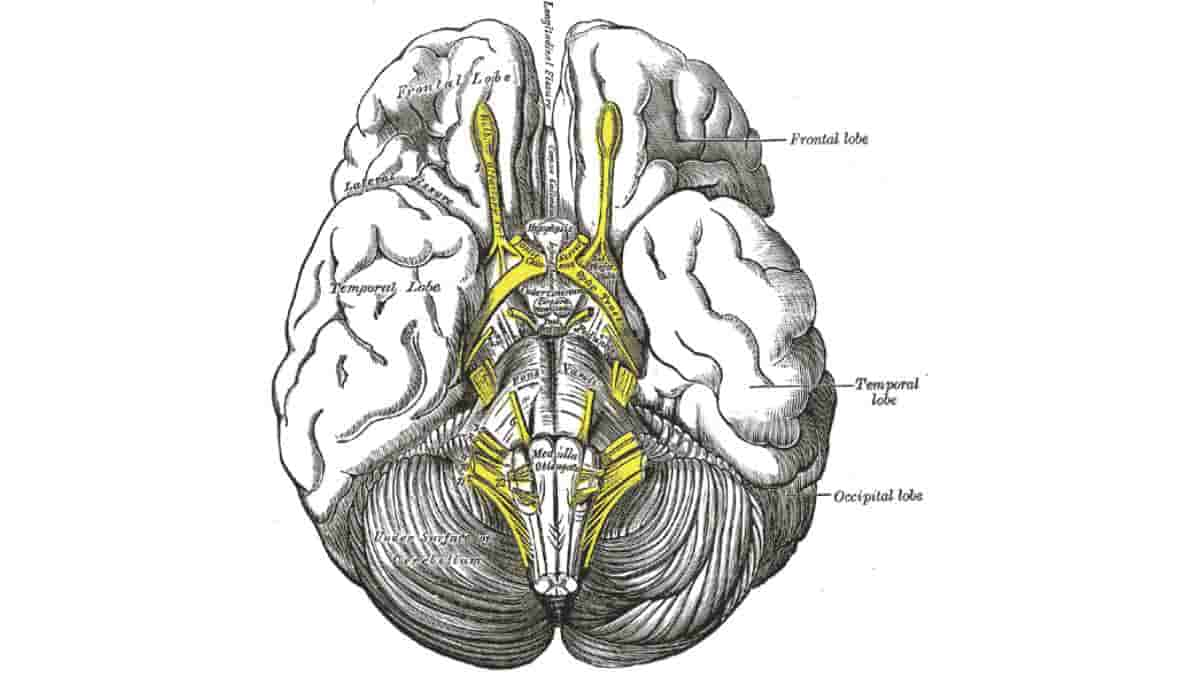
The optic chiasm, also known as the optic chiasma, is the region of the brain where the optic nerves from the right and left eyes intersect. It is situated at the base of the brain, directly beneath the hypothalamus.
The optic nerves of the left and right eyes meet in the body’s midline, ventral to the brain, in all vertebrates. In a number of vertebrates, however, the left optic nerve traverses the right optic nerve without fusing with it.
In vertebrates with a substantial overlap of the visual fields of the two eyes, such as the majority of mammals and birds, as well as amphibians and reptiles such as chameleons, the two optic nerves converge at the optic chiasm.
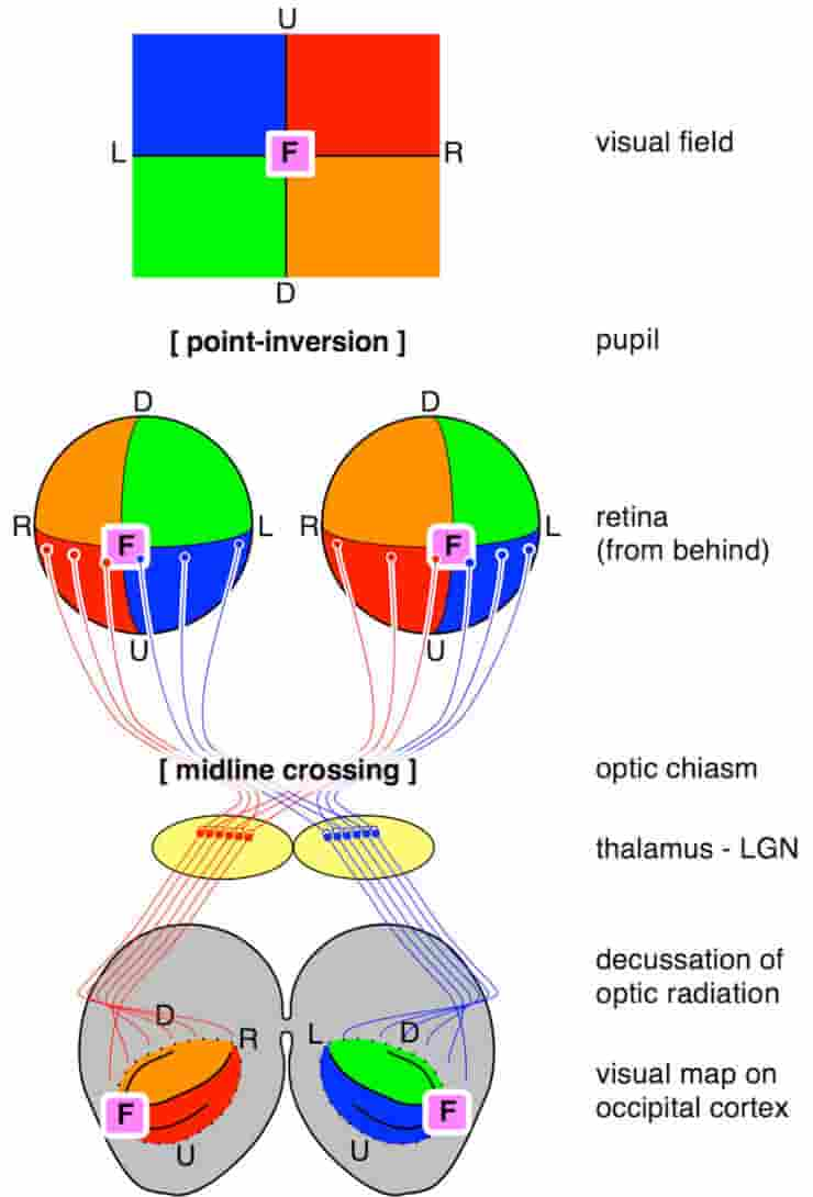
A point-inversion (mirroring) occurs in the pupil, so that U,D as well as L,R are reversed. A (partial) midline crossing takes place in the optic chiasm, and a final mirroring takes place by the decussation of the optic radiation. The final “image” is upside-down, and the left and right hemifields are on the contralateral hemisphere. U=up; D=down; L=left; R=right; F=fovea.
Credit: Marc de Lussanet CC-BY
In a merged optic chiasm, some nerve fibers do not cross the midline but instead, proceed toward the ipsilateral optic tract. By this partial crossing, the portion of the visual field shared by both eyes is merged, allowing stereopsis to process binocular depth perception.
In such cases of partial crossing, the optic nerve fibres on the medial sides of each retina (which correspond to the lateral side of each visual hemifield because the image is inverted) cross to the opposite side of the body’s midline. Inferonasal retinal fibers are associated with the anterior portion of the optic chiasm, while superonasal retinal fibers are associated with the posterior portion.
This partial crossing over of optic nerve fibers at the optic chiasm permits the visual cortex to receive the identical hemispheric visual field from both eyes. These monocular visual signals are superimposed and processed by the visual cortex to generate binocular and stereoscopic vision.
Consequently, the right cerebral hemisphere processes the left visual hemifield, whereas the left cerebral hemisphere processes the right visual hemifield.
Optic Chiasm Development
During development, the crossing of the optic nerves is primarily guided by cues including netrin, slit, semaphorin, and ephrin; and by morphogens including sonic hedgehog (Shh) and Wnt. This navigation is mediated by the neuronal growth cone, a structure that responds to stimuli from ligand-receptor signaling systems by activating pathways that induce cytoskeleton changes.
Retinal ganglion cell (RGC) axons leaving the eye through the optic nerve are blocked from exiting the developing pathway by Slit2 and Sema5A inhibition, expressed bordering the optic nerve pathway. Ssh expressed at the central nervous system midline inhibits crossing prior to the chiasm, where it is downregulated. As they approach the chiasm site, the orientation of RGC axons changes from retinotopic to sheet-like.
Most RGC axons cross the midline at the ventral diencephalon and continue to the contralateral superior colliculus. The number of axons that do not cross the midline and project ipsilaterally depends on the degree of binocular vision of the animal (3% in mice and 45% in humans do not cross).
References:
- Herrera, E; Erskine, L; Morenilla-Palao, C (2019). Guidance of retinal axons in mammals. Seminars in Cell & Developmental Biology. 85: 48–59. doi:10.1016/j.semcdb.2017.11.027
- Nieuwenhuys, R.; Donkelaar, H.J.; Nicholson, C.; Smeets, W.J.A.J.; Wicht, H. (1998). The central nervous system of vertebrates. New York: Springer. ISBN 9783642621277
- Stephen, Polyak (1957). The vertebrate visual system. Chicago: Chicago Univ. Press
Top image: Brain seen from below, with the optic chiasm seen in yellow in the centre. Credit: Henry Gray (1918) Anatomy of the Human Body, Plate 724.