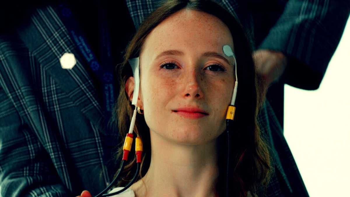
Researchers at Imperial College London are leading the development and testing of a novel brain stimulation technique that may offer an alternative therapy for diseases of the brain like Alzheimer’s and the memory loss that goes along with it. The non-invasive technique, known as temporal interference (TI), delivers electrical fields to the brain via electrodes placed on the patient’s scalp and head.
Researchers were able to stimulate the hippocampus without affecting the surrounding areas by targeting the overlapping electrical fields. Such a procedure previously required surgery to implant electrodes into the brain.
A team from the UK Dementia Research Institute (UK DRI) at Imperial and the University of Surrey successfully tested the approach on 20 healthy volunteers for the first time.
“Until now, if we wanted to electrically stimulate structures deep inside the brain, we needed to surgically implant electrodes which of course carries risk for the patient, and can lead to complications,”
said Dr. Nir Grossman, from the Department of Brain Sciences at Imperial College London.
Temporal Interference Improved Memory
The research team is now conducting a clinical trial in people with early-stage Alzheimer’s disease, where they hope temporal interference could be used to improve symptoms of memory loss. Their preliminary findings indicate that memory function improved when healthy adults performed a memory task while receiving TI stimulation.
With our new technique we have shown for the first time, that it is possible to remotely stimulate specific regions deep within the human brain without the need for surgery. This opens up an entirely new avenue of treatment for brain diseases like Alzheimer’s which affect deep brain structures,”
Dr. Grossman said.

Concept of TI hippocampal stimulation: a, Two current sources I1 and I2 are applied simultaneously via electrically isolated pairs of scalp electrodes (orange and green) at kHz frequencies f1 and f2, with a small frequency difference Δf = f1 − f2 within the range of neural activity. The currents generate oscillating electric fields E\bf E1(t) and E\bf E2(t) inside the brain (orange and green arrows, respectively). Superposition of these fields, E\bf E1(t) + E\bf E2(t), results in an envelope amplitude that is modulated periodically at Δf. The peak amplitude of the envelope modulation can be localized in deep brain structures such as the hippocampus (highlighted in red). b, Schematic of electrode configuration targeting the left hippocampus. Electrodes e1 and e2 formed one electrode pair (orange) and electrodes e3 and e4 another (green), corresponding to I1 and I2 in a. e1 and e3 were located at nasion plane of the left hemisphere, symmetrically above the anterior–posterior midline of the hippocampus (5 cm distance between electrode centers). e2 and e4 were located at a plane above the eyebrow on the right hemisphere (approximately 16 cm distance between electrode centers). Electrodes were 1.5 cm × 1.5 cm square with rounded corners for ex vivo and in vivo experiments and circular 2 cm diameter for computational modeling. c, Illustration of steering of the TI stimulation locus along the hippocampal longitudinal axis. TI stimulation with 1:1 current ratio (‘TI 1:1’) and stimulation locus in the middle region (left); TI stimulation with 1:3 current ratio (‘TI 1:3’) and locus in the anterior region (right). By reducing the current amplitude in one electrode pair and increasing it in the second while keeping the current sum fixed, the stimulation locus can be steered toward the electrode pair with the smaller current amplitude14. Computation of TI stimulation locus in a human anatomical model: d, Schematic of the ROIs in the left (stimulated) hippocampus and its overlying cortex; Ant, anterior; Mid, middle; Post, posterior. e, Left: fields’ envelope modulation amplitude. Right: fields’ absolute amplitude; for the ROIs shown in d. Values are median ± s.d. normalized to the hippocampal value here and thereafter (n indicates number of voxels (nvox) per ROI: Cortex (Crtx), Crtx Ant 48,103, Crtx Mid 43,247, Crtx Post 42,656, Hippocampus 50,349). For whole-brain electric field modeling, see Supplementary Fig. 1a. Note that the cortex ROIs are more heterogeneous than the hippocampus as these include gray matter with different folding and white matter tissue. f, Envelope modulation amplitude in hippocampal ROIs (for ROI schematic, see Fig. 2b) during TI 1:1 and TI 1:3 stimulations (n indicates nvox: Ant 22,651, Mid 17,718, Post 9,980); for additional current ratios, see Supplementary Fig. 1. Measurement of TI stimulation locus in a human cadaver (I1 = 2 kHz, 1 mA; I2 = 2.005 kHz, 1 mA): g, Left: CT head image with intracranial electrode leads a, b and c implanted in the left mesial temporal lobe. Each electrode consisted of 15 electrode contacts; black contour, approximate location of the left hippocampus; orange and green stimulation electrodes. Middle: amplitudes of the envelope modulation in the left (stimulated) hippocampus and its overlying cortex showing higher envelope amplitude at the hippocampus (LMM, two-sided paired t-test, t(2) = −5.515, P = 0.0345, n = 3 electrodes). Right: absolute amplitudes in the left hippocampus and overlying cortex, showing higher absolute amplitude in the overlying cortex (LMM, two-sided paired t-test, t(2) = 7.051, P = 0.0195). Dots represent individual electrodes. See Supplementary Table 1 for full statistics and Supplementary Fig. 2 for additional amplitude maps. h, Envelope modulation ratio versus depth for electrode b, showing increasing envelope modulation with depth. i, Anterior (Ant) to posterior (Post) envelope modulation amplitude for the TI 1:1 and TI 1:3 conditions, showing higher Ant/Post amplitudes for the TI 1:3 condition (two-sided paired t-test, t(7) = −7.765, P = 1.204 × 10⁻⁴, n = 8 hippocampal electrode contacts); envelope modulation amplitudes in the anterior electrode a were normalized to the posterior electrode c. Dots represent individual contacts in the hippocampal region and are color coded by depth (cold colors for more superficial contacts and warmer colors for deeper contacts). Asterisks identify significant differences, P < 0.05. Bar plots show median ± s.d.
Credit: Nature Neuroscience (2023). DOI:10.1038/s41593-023-01456-8
TI was first described by the team at Imperial College London in 2017 and shown to work in principle in mice. This new research demonstrates for the first time that TI is capable of stimulating deep brain regions in humans.
According to the researchers, this could have broad applications and will enable scientists to stimulate different deep brain regions to discover more about their functional roles, accelerating the discovery of new therapeutic targets.
Hippocampal Focus
The study used measurements of brains that had already died to show that electric fields could be focused on the hippocampus from a distance. The hippocampus is a deep, curved part of the brain that is very important for learning and remembering things.
The team then applied the TI stimulation to healthy volunteers while they were memorizing pairs of faces and names — a process heavily dependent on the hippocampus. Researchers used functional magnetic resonance imaging (fMRI) to show that TI changed the activity in the hippocampus that was caused by the memory task.
Finally, the researchers repeated the procedure for a longer period of 30 minutes. This showed that TI stimulation during the task lead to improved memory accuracy.
Better Individualized Strategies
Dr. Ines Violante, from the University of Surrey and an Honorary Research Fellow at Imperial, and the study’s first author, stated that the ability to target deep brain areas using a non-invasive approach selectively is very exciting because it provides a tool to investigate how the human brain functions and opens the door to clinical applications.
“The combination of non-invasive imaging and brain stimulation will help us unravel the processes that support our cognitive functions, such as memory and learning. Knowledge of these processes and how they can be altered is essential to develop better individualized strategies to treat or delay the onset of diseases,”
he said.
We hope this work will help to scale up the availability of deep brain stimulation therapies by drastically reducing cost and risk. We are now testing whether repeated treatment with the stimulation over the course of a number of days could benefit people in the early stages of Alzheimer’s. We hope that this will restore normal brain activity in the affected areas, which could improve symptoms of memory impairment,”
Added Dr. Grossman.
This study was released concurrently with a second article led by researchers at Switzerland’s École polytechnique fédérale de Lausanne (EPFL), which independently validated the technology.
In their study, which was also published in Nature Neuroscience, EPFL researchers used TI technology to focus on stimulating a different part of the brain called the striatum. This helped healthy volunteers improve their motor memory.
Abstract:
Deep brain stimulation (DBS) via implanted electrodes is used worldwide to treat patients with severe neurological and psychiatric disorders. However, its invasiveness precludes widespread clinical use and deployment in research. Temporal interference (TI) is a strategy for non-invasive steerable DBS using multiple kHz-range electric fields with a difference frequency within the range of neural activity. Here we report the validation of the non-invasive DBS concept in humans. We used electric field modeling and measurements in a human cadaver to verify that the locus of the transcranial TI stimulation can be steerably focused in the hippocampus with minimal exposure to the overlying cortex. We then used functional magnetic resonance imaging and behavioral experiments to show that TI stimulation can focally modulate hippocampal activity and enhance the accuracy of episodic memories in healthy humans. Our results demonstrate targeted, non-invasive electrical stimulation of deep structures in the human brain.
References:
- Violante, I.R., Alania, K., Cassarà, A.M. et al. Non-invasive temporal interference electrical stimulation of the human hippocampus. Nature Neuroscience (2023). DOI: 10.1038/s41593-023-01456-8
- Wessel, M.J., Beanato, E., Popa, T. et al. Noninvasive theta-burst stimulation of the human striatum enhances striatal activity and motor skill learning. Nature Neuroscience (2023). DOI: 10.1038/s41593-023-01457-7
Top image credit: Imperial College London