Neuroscience is in a state of tremendous flux. Neurons and glial cells, the two main cell types that make up the brain, have a hybrid cell halfway between these two categories.
For as long as neuroscience has existed, it has been recognized that the brain functions primarily through the neurons and their ability to rapidly elaborate and transmit information through their networks.
But now, a previously unknown type of cell that is essential for brain function has been identified by a research team from the University of Lausanne (UNIL) and the Wyss Center. These cells of a novel order, hybrid in composition and function between the two known types of brain cells (neurons and glial cells), are found in multiple regions of the mouse and human brain.
The research shows that these cells improve memory and brain control of movement, as well as help to prevent epileptic seizures.
Debate Finally Ended
Glial cells perform a variety of structural, energetic, and immune functions, in addition to stabilizing physiological constants, to aid neurons in their duties. Some of these glial cells, known as astrocytes, are located in close proximity to synapses, the contact sites where neurotransmitters are released to transmit information between neurons.
Therefore, neuroscientists have long hypothesized that astrocytes may participate in synaptic transmission and information processing. The studies conducted to date to demonstrate this, however, have produced contradictory results and have not yet attained a scientific consensus.

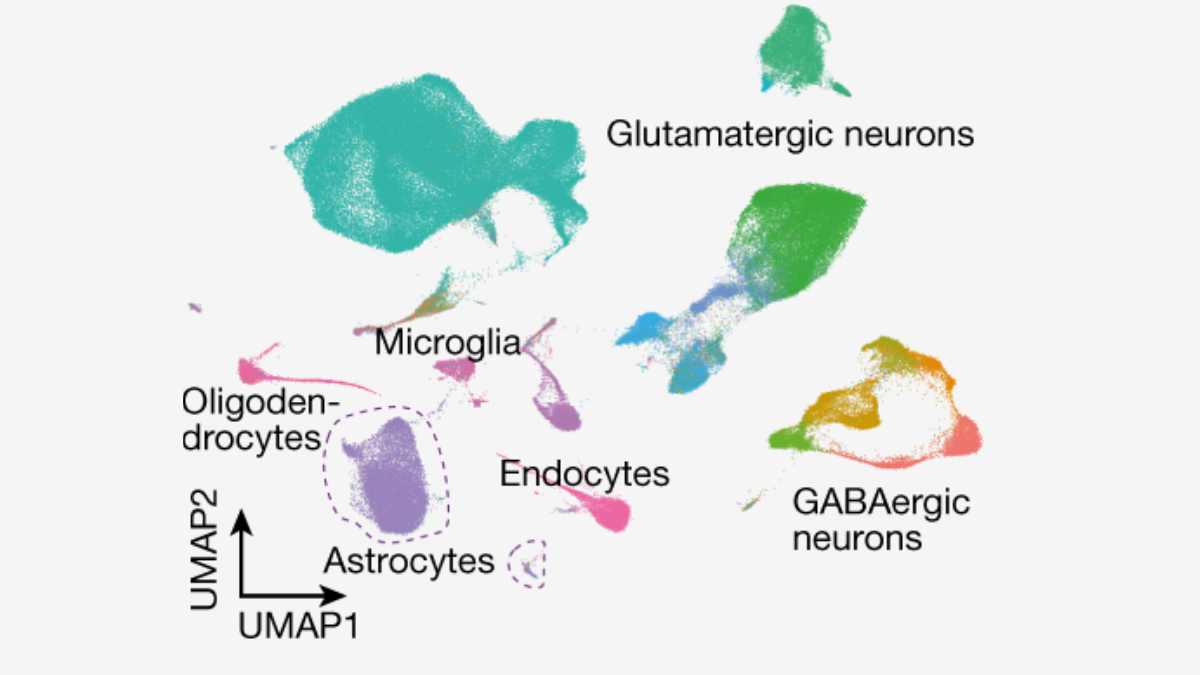
Credit: Nature. 2023;10.1038/s41586-023-06502-w CC-BY
Neuroscientists from the UNIL’s Faculty of Biology and Medicine and the Wyss Center for Bio and Neuroengineering in Geneva put an end to years of debate by identifying a new cell type with the characteristics of an astrocyte and expressing the molecular machinery required for synaptic transmission.
Glutamatergic Vesicle Proteins
To confirm or refute the hypothesis that astrocytes, like neurons, can release neurotransmitters, researchers examined the molecular composition of astrocytes using contemporary molecular biology techniques. Their objective was to identify traces of the apparatus required for the rapid secretion of glutamate, the principal neurotransmitter utilized by neurons.

Right, the stimulation protocol used for drug applications. Ten-millisecond puff applications were performed six times, one every 20 s, during 120 s imaging acquisitions.
Credit: Nature. 2023;10.1038/s41586-023-06502-w CC-BY
Researchers were able to demonstrate the presence of transcripts of the vesicular proteins, VGLUT, responsible for filling neuronal vesicles specific for glutamate release, in cells with an astrocytic profile. This was due to the precision afforded by single-cell transcriptomics techniques. These transcripts were discovered in mouse cells and appear to be preserved in human cells.
“We also identified other specialized proteins in these cells, which are essential for the function of glutamatergic vesicles and their capacity to communicate rapidly with other cells,”
said Ludovic Telley, Assistant professor at UNIL, co-director of the study.
Advanced In-vivo Imaging
Neuroscientists then investigated whether these composite cells were functional, i.e., capable of releasing glutamate at a rate comparable to synaptic transmission. To accomplish this, the research team employed a cutting-edge imaging technique (single-wavelength intensity-based glutamate-sensing fluorescent reporters) that allowed for the visualization of glutamate released by vesicles in the brain tissues of living rodents.
“We have identified a subgroup of astrocytes responding to selective stimulations with rapid glutamate release, which occurred in spatially delimited areas of these cells reminiscent of synapses,”
said study co-director Andrea Volterra, honorary professor at UNIL.
This glutamate release also regulates synaptic transmission and neuronal circuitry. The research team was able to illustrate this by inhibiting VGLUT expression in hybrid cells.
They are cells that regulate neuronal activity by controlling the level of communication and excitation of neurons, explained Roberta de Ceglia, first author of the study and senior researcher at UNIL.
And, without this functional machinery, the study demonstrates that long-term potentiation, a neural process involved in the mechanisms of memory, is impaired and that mice have impaired memories.
Brain Pathology Involvement
This discovery has implications for neurological conditions. The research team demonstrated effects on memory consolidation by disrupting glutamatergic astrocytes specifically.
However, they also discovered associations with diseases such as epilepsy, whose seizures were exacerbated. Lastly, the study demonstrates that glutamatergic astrocytes play a role in the modulation of brain circuits involved in the control of movement and may offer potential therapeutic targets for Parkinson’s disease.
“In between neurons and astrocytes, we now have a new kind of cell at hand. Its discovery opens up immense research prospects. Our next studies will explore the potential protective role of this type of cell against memory impairment in Alzheimer’s disease, as well as its role in other regions and pathologies than those explored here,”
said Andrea Volterra.
The authors anticipate future studies to generate Central Nervous System-wide maps that will aid in defining the overall distribution of glutamatergic astrocytes and their full spectrum of actions. These could help us understand why this atypical astrocytic population exists, how it anatomically and functionally integrates into CNS circuits, and whether and how its altered properties contribute to specific pathological CNS conditions.
Reference:
- de Ceglia R, Ledonne A, Litvin DG, et al. Specialized astrocytes mediate glutamatergic gliotransmission in the CNS. Nature. 2023; 10.1038/s41586-023-06502-w. doi: 10.1038/s41586-023-06502-w
Last Updated on December 21, 2023