A new study from the Institute of Psychiatry, Psychology, and Neuroscience has discovered a link between a decrease in gray matter in the brain and early onset psychosis (EOP).
The research, the largest brain imaging study ever conducted in EOP, offers unparalleled amounts of data regarding the illness. It demonstrates that, in comparison to other mental health illnesses, patients with EOP have a lower volume of gray matter in practically all regions of their brain.
Researchers hope that this detailed mapping can be used to aid in future diagnosis and to track the effects of treatment in EOP patients.
Left Median Cingulate
Early onset psychosis occurs before the age of 18 and happens during a vital phase of brain development. Individuals with the condition are more prone to have severe and long-lasting symptoms that respond poorly to treatment. Despite this, EOP research has been constrained in terms of sample size and statistical power.
The study is an international collaboration, combining brain scans from Norway, Spain, Canada, Italy, Australia, and the United Kingdom, with 482 people with EOP being compared to 469 healthy controls.
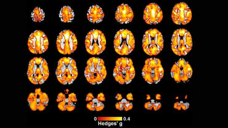
An analysis of the data revealed that individuals with EOP had lower gray matter volumes in almost all regions of the brain compared to healthy controls, with a notable effect in the left median cingulate — an area of the brain associated with the formation and processing of emotions, learning, and memory.
“Early onset psychosis can have a devastating impact on a person’s life and well-being, but our understanding of the illness is still sadly relatively limited. This study, the largest neuroimaging analysis of EOP to date, used newly developed technologies to combine scans from different sites to examine hundreds of thousands of data points measuring volume in the brain,”
said lead author Dr. Matthew Kempton, Reader in Neuroimaging Psychiatry at King’s IoPPN.
Specially Created Software
The researchers discovered that persons with EOP have less gray matter in nearly all regions of their brains than people who do not have the condition. This precise map will ideally serve as the foundation for future study, as it might be used as a diagnostic tool and even to track the efficacy of therapies.
Further analysis of the data revealed that people who developed early onset psychosis later in life had lower gray matter volumes in a number of small brain regions than those who developed it earlier.
“Gray matter’s primary purpose is to process information in the brain and plays a significant role in day-to-day functions like memory, emotions and movement. This study used specially created software (ENIGMA-VBM) developed at King’s that can accurately map where there have been local increases and decreases in brain volume. It’s allowed our team to process significantly more data and has meant that our sample reflects brain scans from many parts of the world. The effectiveness of this software means we’re now investigating the brains of those with several other disorders,”
Shuqing Si, the study’s first author from King’s IoPPN, said.
Abstract
Introduction: Regional gray matter (GM) alterations have been reported in early-onset psychosis (EOP, onset before age 18), but previous studies have yielded conflicting results, likely due to small sample sizes and the different brain regions examined. In this study, we conducted a whole brain voxel-based morphometry (VBM) analysis in a large sample of individuals with EOP, using the newly developed ENIGMA-VBM tool.
Methods: 15 independent cohorts from the ENIGMA-EOP working group participated in the study. The overall sample comprised T1-weighted MRI data from 482 individuals with EOP and 469 healthy controls. Each site performed the VBM analysis locally using the standardized ENIGMA-VBM tool. Statistical parametric T-maps were generated from each cohort and meta-analyzed to reveal voxel-wise differences between EOP and healthy controls as well as the individual-based association between GM volume and age of onset, chlorpromazine (CPZ) equivalent dose, and other clinical variables.
Results: Compared with healthy controls, individuals with EOP showed widespread lower GM volume encompassing most of the cortex, with the most marked effect in the left median cingulate (Hedges’ g = 0.55, p = 0.001 corrected), as well as small clusters of lower white matter (WM), whereas no regional GM or WM volumes were higher in EOP. Lower GM volume in the cerebellum, thalamus and left inferior parietal gyrus was associated with older age of onset. Deficits in GM in the left inferior frontal gyrus, right insula, right precentral gyrus and right superior frontal gyrus were also associated with higher CPZ equivalent doses.
Conclusion: EOP is associated with widespread reductions in cortical GM volume, while WM is affected to a smaller extent. GM volume alterations are associated with age of onset and CPZ equivalent dose but these effects are small compared to case-control differences. Mapping anatomical abnormalities in EOP may lead to a better understanding of the role of psychosis in brain development during childhood and adolescence.
Reference:
- Si, S., Bi, A., Yu, Z. et al. Mapping gray and white matter volume abnormalities in early-onset psychosis: an ENIGMA multicenter voxel-based morphometry study. Mol Psychiatry (2024). Doi: 10.1038/s41380-023-02343-1
Image: Regions showing lower Gray Matter volume in individuals with Early Onset Psychosis compared to Healthy Controls, Credit: Mol Psychiatry (2024). Doi: 10.1038/s41380-023-02343-1