A Korean study team has established, for the first time, a link between depression and taurine concentrations in the hippocampus, the part of the brain responsible for memory and learning functions. This discovery presents a chance to emphasize the role and significance of taurine in future depression prevention, diagnosis, and therapy.
Using ultra-high magnetic field 7T human MRI (7T MRI), researchers (Drs. Youngkyu Song, Jee-Hyun Cho, and Chaejoon Cheong) from the Korea Basic Science Institute (KBSI) Biochemical Analysis Team confirmed that taurine concentration was significantly lower in the hippocampus of young females suffering from depression.
Dr. Hyungjun Kim of the Korea Institute of Oriental Medicine (KIOM) and Prof. Jin-Hun Sohn of Chungnam National University (CNU) led research teams that conducted the study, which compared two groups of female participants — 36 patients with major depressive disorder and 40 healthy females. Participants ranged in age from 19 to 29.
Rates of Depression Increase Doubled
Depression causes significant harm and loss, not just to the individual, but also to society and the economy. According to the World Health Organization (WHO), more than 260 million people worldwide suffer from depression, and more than 800,000 individuals commit suicide each year.
In Korea, the increase in depression among young people is notable. Of the total 1,000,744 patients with depression, 185,942 individuals in their 20s accounted for the largest demographic, and the rate of increase has more than doubled in five years.
Because it can precisely scan particular places in the body and gather a range of quantitative data, magnetic resonance imaging (MRI) is widely employed in brain disorder research. Previous MRI research on depression concentrated on revealing changes in metabolites mostly in the cerebral cortex area, near the brain’s border.
Hippocampus Metabolites
This study is the first to disclose the relationship between metabolites and depression in the hippocampus, located inside the brain.
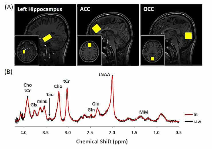
The research team conducted an analysis to identify substances that are strongly associated with depression. To achieve this, they measured and compared the concentrations of seven metabolites found in the frontal, occipital, and hippocampus regions of young women: taurine, choline, creatine, glutamine, glutamate, myo-inositol, and N-acetyl aspartate.
When performing MRI scans, there are technical limitations in measuring metabolite concentration in the hippocampus due to its location in the brain. Also, it is particularly difficult to obtain a magnetic resonance spectroscopy (MRS) signal for taurine because it has a low concentration compared to other metabolites.
The researchers were able to uncover subtle differences in taurine signals in the hippocampus of the patient and control groups by using a 7T MRI scanner, which is very sensitive and has a high resolution, along with a sLASER pulse sequence that is designed to reduce chemical shift displacement errors.
Taurine and Depression
It was also possible to precisely measure metabolite concentrations while taking into account the different ways that white matter, gray matter, and cerebrospinal fluid (CSF) are distributed in each person. These metrics are expected to be used in customized brain disease studies in the future, according to individual characteristics.
“This study will promote research on the role of taurine in the hippocampus and its relationship with depression, and contribute to the pathogenesis research and diagnosis development of depression,”
said the research team leader, Dr. Jee-Hyun Cho.
“By using KBSI’s cutting-edge research equipment, we plan to conduct follow-up research on changes of taurine concentrations in the brain via long-term observation of depression patients, as well as the effect of taurine intake as a treatment for depression,”
she added.
Limitations
The Korea Basic Science Institute study team provided the basic research hypothesis of a link between depression and taurine concentration in the hippocampus, measured brain metabolites using 7 Tesla MRI, and analyzed the resultant data. The KIOM and Chungnam National University research teams assisted in the recruitment of depression patients and healthy control groups, as well as the administration of psychological tests and clinical interviews and the management of demographic data.
The small number and range of participants is one of the study’s limitations. Participants in future trials should be older and male.
The participants of this study were all young women with mean age, of 22.02 and 22.62 years for major depressive disorder and healthy controls, respectively. The study focused on women because they are more vulnerable to MDD, almost twice as likely as men.
Abstract
Background: Major depressive disorder (MDD) is characterized by depressed mood or loss of interest or pleasure. Generally, women are twice as likely as men to have depression. Taurine, a type of amino acid, plays critical roles in neuronal generation, differentiation, arborization and formation of synaptic connections. Importantly, it enhances proliferation and synaptogenesis in the hippocampus. When injected into animals, taurine has an antidepressant effect. However, there is no in vivo evidence to show the association between taurine concentration in the human brain and development of MDD.
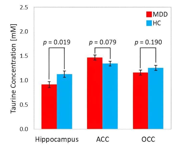
Methods: Forty-one unmedicated young (aged 18-29) women with MDD and 43 healthy controls (HCs) matched for gender and age were recruited in South Korea. The taurine concentration was measured in the hippocampus, anterior cingulate cortex and occipital cortex of the MDD and HC groups using proton magnetic resonance spectroscopy at 7T. Analysis of covariance was used to examine differences in taurine concentration, adjusted for age as a covariate.
Results: The taurine concentration in the hippocampus was lower (F1,75 = 5.729, p = 0.019, Δη2 = 0.073) for MDD (mean (SEM), 0.91 (0.06) mM) than for HC (1.13 (0.06) mM). There was no significant difference in taurine concentration in the anterior cingulate cortex or occipital cortex between the two groups.
Conclusions: This study demonstrates that a lower level of taurine concentration in the hippocampus may be a novel characteristic of MDD.
Reference:
- Youngkyu Song et al. Association between taurine level in the hippocampus and major depressive disorder in young women: a proton magnetic resonance spectroscopy study at 7 Tesla. Biological Psychiatry (2023). DOI: 10.1016/j.biopsych.2023.08.025