The somatic nervous system is a part of the peripheral nervous system, which works in coordination with the central nervous system to control various bodily functions. Together, these systems form the complex neural network responsible for transmitting information and executing essential tasks in the body.
The peripheral nervous system comprises two major divisions: the somatic nervous system and the autonomic nervous system. While the autonomic nervous system manages involuntary processes, such as digestion and heart rate regulation, the somatic nervous system is primarily responsible for voluntary movements and the perception of external stimuli. It consists of afferent and efferent neurons, which facilitate communication between the body’s sensory and motor systems.
Afferent neurons, also known as sensory neurons, carry information from the body’s sensory receptors—including those found in the skin, muscles, and joints—toward the central nervous system. This information is processed and interpreted within the central nervous system, which includes the brain and spinal cord. The central nervous system then sends commands back to the muscles and other structures through efferent neurons, also called motor neurons.
The main functions of the somatic nervous system are:
-
- Gathering sensory information from the body’s external environment
- Relaying sensory information to the central nervous system
- Transmitting motor commands from the central nervous system to the muscles
- Coordinating voluntary movements and responses to external stimuli
Somatic Nervous System Functional Aspects
The somatic nervous system plays a crucial role in voluntary movements. It is responsible for initiating the motor function of skeletal muscles by sending signals from the central nervous system (CNS) to the muscles.
When we decide to move a part of our body, the somatic nervous system is triggered and releases the neurotransmitter acetylcholine at the neuromuscular junction, leading to muscle contraction.
Sensory Input Processing
The somatic nervous system is also involved in sensory function. It processes and transmits sensory information from external stimuli (such as touch, pain, and temperature) through peripheral nerves to the CNS. This sensory input is critical in allowing the body to recognize and adapt to changes in the environment
An important concept within somatic sensory processing is the concept of excitatory and inhibitory signals. Excitatory signals increase the likelihood of generating an action potential, while inhibitory signals reduce this likelihood. This modulation of sensory signals allows the somatic nervous system to effectively process incoming sensory information and respond accordingly.
Reflex Arc Mechanisms
The somatic nervous system also plays a key role in reflex arc mechanisms. These simple pathways are responsible for generating reflexes or reflex actions, automatic and rapid motor responses to specific stimuli that protect the body from harm. Some common examples of reflexes include withdrawing the hand from a hot surface or quickly closing the eye when an object approaches it.
A reflex arc typically involves a sensory receptor, which detects the stimulus, a sensory neuron (afferent), a higher-order integrating center (usually in the spinal cord), a motor neuron (efferent), and an effector, which carries out the motor response. The key feature of a reflex arc is its ability to bypass conscious control, allowing the body to respond quickly and effectively to potential threats to its well-being.
Structural Components
The somatic nervous system consists of sensory neurons that detect and transmit information from sensory receptors to the central nervous system (CNS). Sensory receptors are specialized structures that detect various stimuli, such as temperature, pain, pressure, and proprioception.
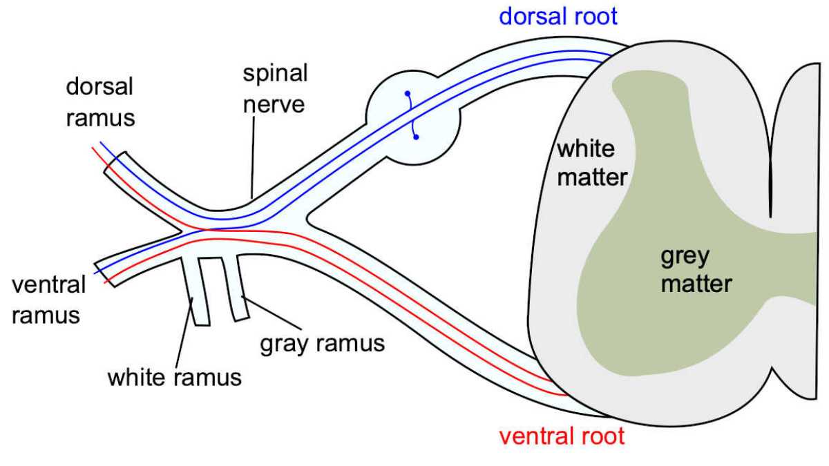
Sensory neurons, also known as afferent neurons, have their cell bodies located in the dorsal root ganglia of the spinal cord, while their axons extend to both the peripheral target receptor and the spinal cord.
Axons of sensory neurons are usually covered with myelin, a layer of insulation that speeds up the conduction of nerve impulses. Dendrites link with sensory receptors and relay information to the cell body, ultimately transmitting the signal through synapses to the spinal cord or directly to the brain via the cranial nerves.
Motor Neurons and Muscles
Motor neurons, also called efferent neurons, transmit signals from the CNS to the skeletal muscles, initiating muscle contraction and movement. These neurons have their cell bodies in the ventral horn of the spinal cord or within the cranial nerve nuclei. Motor neurons communicate directly with the muscles through the neuromuscular junction.
In the neuromuscular junction, the axon terminal of the motor neuron makes contact with the muscle fiber, forming a synapse. The motor neuron releases neurotransmitters, which bind to receptors on the muscle fiber, causing it to contract. This process is essential for the overall function of the somatic nervous system, as it facilitates voluntary movements and reflexes.
Neural Pathways
The nerves of the somatic nervous system are classified according to their location, either in the head or in the spine. There are 12 pairs of cranial nerves that send information to or from the brain stem (the base of the brain where the spinal cord joins).
Neural pathways within the SNS connect the sensory receptors and motor neurons, allowing for the transmission of sensory information and muscle control. Several types of neural pathways exist in the SNS, but most involve sequences of interconnected neurons within the spinal cord and the brain.
For example, a simple reflex arc includes a sensory neuron that detects a stimulus, an interneuron within the spinal cord that processes the information, and a motor neuron that controls the muscle response. More complex pathways may involve higher brain centers in which sensory information is processed, integrated, and used to plan and execute voluntary movements.
Pathology and Disorders
The somatic nervous system is involved in many common conditions related to nerve damage and dysfunction. Infections and toxins can target the somatic nerves leading to various disorders, including peripheral neuropathy.
Peripheral neuropathy often leads to symptoms such as numbness, tingling, and weakness in the affected extremities. This disorder affects the nerves that transmit signals between the brain, spinal cord, and the rest of the body.
One common cause of peripheral neuropathy is diabetes, a condition in which prolonged high blood sugar levels can inflict damage on the nerves. Additionally, autoimmune diseases, such as lupus and rheumatoid arthritis, can lead to inflammation and damage to the somatic nerves. Systemic infections may also contribute to peripheral neuropathy, as they can invade and harm nervous tissue.
Infections and toxins can have direct effects on the somatic nervous system. For example, the bacterial infection, Lyme disease, may lead to peripheral neuropathy. Exposure to certain toxins from certain medications, chemicals, or heavy metals can result in nerve damage, causing somatic symptoms like numbness and involuntary movements.
A variety of physical trauma and injuries can also lead to somatic nerve damage, such as nerve compression or pressure. Carpal tunnel syndrome is a familiar example of peripheral neuropathy caused by pressure on a specific somatic nerve. In severe cases, physical nerve damage can lead to more extensive issues, such as tissue damage or muscle atrophy.
Another condition affecting the SNS is Trigeminal neuralgia, also known as Fothergill illness, tic douloureux, or trifacial neuralgia, is a chronic pain disorder that affects the trigeminal nerve, which is responsible for feeling in the face as well as motor tasks such as biting and chewing. It is classified as neuropathic pain.
It is regarded as one of the most painful medical disorders, and frequently leads to depression and suicide. The specific cause is unknown, but it is thought to be a loss of trigeminal nerve myelin. This could be caused by a blood vessel compressing the nerve as it exits the brain stem, multiple sclerosis, stroke, or trauma.
References:
- Brookhart, J M. Somatic Functions of the Central Nervous System. Annual Review of Physiology 1954 16:1, 325-348
- Cracchiolo, Marina et al. Bioelectronic medicine for the autonomic nervous system: clinical applications and perspectives. Journal of neural engineering vol. 18,4 10.1088/1741-2552/abe6b9. 17 Mar. 2021, doi:10.1088/1741-2552/abe6b9
- Cruccu G, Di Stefano G, Truini A (August 2020). Ropper AH (ed.). Trigeminal Neuralgia. The New England Journal of Medicine. 383 (8): 754–762. doi:10.1056/NEJMra1914484
- Hoehn-Saric, R, and D R McLeod. The peripheral sympathetic nervous system. Its role in normal and pathologic anxiety. The Psychiatric clinics of North America vol. 11,2 (1988): 375-86
- McLeod, J.G. and Tuck, R.R. (1987), Disorders of the autonomic nervous system: Part 1. Pathophysiology and clinical features. Ann Neurol., 21: 419-430.
- Purves, Dale (2011). Neuroscience (5th ed.). Sunderland, Mass.: Sinauer. ISBN 9780878936953
- Rea, Paul (2014). Introduction to the Nervous System. Clinical Anatomy of the Cranial Nerves. Academic Press. ISBN 978-0-12-800898-0
- Saladin, Kenneth (2024). Anatomy & Physiology: The Unity of Form and Function (10th ed.). New York, NY: McGraw Hill. ISBN 9781266041846
- Westfall TC, Westfall DP. Neurotransmission: The Autonomic and Somatic Motor Nervous Systems. In: Brunton LL, Chabner BA, Knollmann BC. eds. Goodman & Gilman’s: The Pharmacological Basis of Therapeutics, 12e. McGraw Hill; 2015. Accessed December 14, 2023.
Top image: formation of the spinal nerve from the dorsal and ventral roots. Credit: Mysid, original by Tristanb. CC-BY WM
Last Updated on April 5, 2024