Our pupils reflect our level of arousal: they dilate when we are tense, stressed, or even panicked, and constrict when we relax. A nucleus in the pons of the brainstem known as locus coeruleus, which measures roughly 15 millimeters, is crucial to this. It governs our level of arousal via the neurotransmitter noradrenaline and is located deep within the brain.
Up until now, it had been unclear whether information about pupil size can also be used to volitionally influence the arousal centers in the brain. A new study from ETH Zurich has revealed that this is indeed possible.
“With the right biofeedback, people can better learn to control their state of arousal through mental relaxation and activation techniques,”
explained Nicole Wenderoth, Professor of Neural Control of Movement at ETH Zurich.
The study’s findings open up new avenues for treating stress and anxiety disorders.
Pupil Size Regulation
The researchers first taught a group of 27 subjects to control their pupil size volitionally in order to investigate the relationship between pupil size and the brain’s state of arousal.
Using mental relaxation and activation techniques, such as focusing on breathing and visualizing stressful or threatening situations, the subjects were asked to alternately constrict and dilate their pupils. They sat in front of a screen with an eye tracker that recorded how well they were doing.
The researchers displayed this feedback to some of the subjects on the screen in the form of a circle. The individuals were told by a smaller circle that their pupils were narrowing, which was an indication of relaxation. A growing circle, on the other hand, suggested dilated pupils and increased arousal.
Individuals who got real-time input on their pupil size were shown to be better able to modulate their state of arousal and pupil size. Subjects were able to identify the relaxation and activation approaches that worked best for them, thanks to the pupil feedback.
By contrast, the control group either received a false feedback signal unrelated to their own pupil size or were instructed to focus purely on the use of mental strategies.
Locus Coeruleus Activity
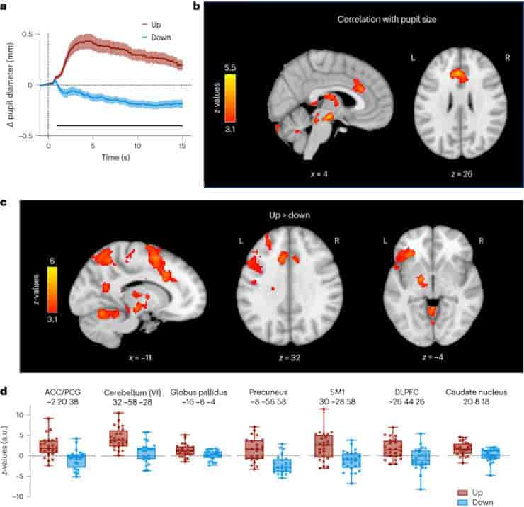
a, Changes in pupil size averaged across all participants for Up (red) and Down (blue) trials showing successful self-regulation of pupil size during whole-brain fMRI recordings. The solid black line at the bottom indicates a cluster of significantly higher baseline-corrected pupil sizes during Up than during Down trials (two-tailed SPM1D paired-samples t-test; P = 0; z* = 3.38). b, Whole-brain maps showing brain regions where BOLD activity correlates with pupil size changes throughout the fMRI runs (GLM). c, Whole-brain maps depicting brain regions that showed significant activation during Up (as compared to Down) trials (GLM). All activation maps in b and c are thresholded at z > 3.1 and FWE-corrected for multiple comparisons using a cluster significance level of P < 0.05. d, Estimated BOLD response represented by z-values for Up vs rest and Down vs rest extracted from the peak voxel of each significant cluster shown in c (n = 24). Boxplots indicate median (centre line), 25th and 75th percentiles (box), and maximum and minimum values (whiskers).
Credit: Nat Hum Behav (2023). doi: 10.1038/s41562-023-01729-z
The experiment was then repeated, but this time the researchers used magnetic resonance imaging to record the subjects’ brain activity.
“We saw that volitional changes in pupil size are actually accompanied by changes in activity in brainstem regions that regulate the brain’s state of arousal,”
said postdoctoral researcher Sarah Meissner.
A look at the subjects’ heart rates revealed that the change in pupil size also physically relaxed or roused them. The pulse of those who were able to better control their pupils as a result of the feedback fell or rose more at the end of the training compared to the beginning of the training and more than the pulse of the control group.
The researchers’ method is applicable to commercially available VR headsets that provide real-time pupil size feedback.
“Our goal is for people to learn to control their pupils in a playful way and, in doing so, find out which relaxation or activation techniques work best for them,”
Wenderoth said. Mindmetrix, an ETH spin-off, has already been established to bring this technology to market.
Abstract
The brain’s arousal state is controlled by several neuromodulatory nuclei known to substantially influence cognition and mental well-being. Here we investigate whether human participants can gain volitional control of their arousal state using a pupil-based biofeedback approach. Our approach inverts a mechanism suggested by previous literature that links activity of the locus coeruleus, one of the key regulators of central arousal and pupil dynamics. We show that pupil-based biofeedback enables participants to acquire volitional control of pupil size. Applying pupil self-regulation systematically modulates activity of the locus coeruleus and other brainstem structures involved in arousal control. Furthermore, it modulates cardiovascular measures such as heart rate, and behavioural and psychophysiological responses during an oddball task. We provide evidence that pupil-based biofeedback makes the brain’s arousal system accessible to volitional control, a finding that has tremendous potential for translation to behavioural and clinical applications across various domains, including stress-related and anxiety disorders.
Reference:
- Meissner, S.N., Bächinger, M., Kikkert, S. et al. Self-regulating arousal via pupil-based biofeedback. Nat Hum Behav (2023). doi: 10.1038/s41562-023-01729-z
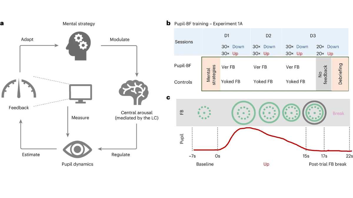
Top Image: a, Participants apply mental strategies that are believed to modulate the brain’s arousal levels mediated by nuclei such as the LC. Pupil size was measured by an eye tracker and fed back to the participant via an isoluminant visual display. b, In experiment 1A, healthy volunteers were informed about potential mental strategies of arousal regulation and then participated in 3 days (D1, D2, D3) of upregulation and downregulation trainings (30 trials each) while receiving either veridical pupil feedback (Ver FB) (pupil-BF group) or visually matched input/yoked feedback (Yoked FB) (control groups I and II). At the end of day 3, all participants performed 20 Up and 20 Down trials without receiving any feedback and were debriefed on which strategies they have used. c, Example trial of the experiment. Each trial consisted of (1) 7 s baseline measurements, (2) 15 s modulation phase where the pupil-BF group sees a circle that dynamically changes its diameter as a function of pupil size (veridical feedback), (3) 2 s of color-coded post-trial performance feedback (green, average circle size during modulation; black, maximum (Up) or minimum (Down) circle size during modulation) and (4) 5 s break. The upper panel shows an example of what participants would see on their screen, while the red line in the lower panel indicates measured pupil size. Note that the control groups I and II see a circle that changes independently of pupil size but resembles that for a participant in the pupil-BF group. Credit: Nature Human Behavior (2023). DOI:10.1038/s41562-023-01729-z