
According to Weill Cornell Medicine neuroscientists, ocular drift, a very subtle and seemingly random type of eye movement, can be influenced by prior knowledge of the expected visual target, implying a surprising level of cognitive control over the eyes.
The discovery advances scientific understanding of how vision, which is more than just the absorption of incoming retinal signals, is controlled and directed by cognitive processes.
“These eye movements are so tiny that we’re not even conscious of them, and yet our brains somehow can use the knowledge of the visual task to control them,”
said lead author Dr. Yen-Chu Lin, who works in the laboratory of the study’s senior author Dr. Jonathan Victor, the Fred Plum Professor of Neurology at Weill Cornell Medicine.
Ocular Drifts
The majority of studies on cognitive control of eye movement have focused on more obvious movements, such as saccade movements in which the eyes dart across large areas of the visual field. Drs. Lin and Victor, along with their colleagues, investigated ocular drift, or tiny jitters of the eye that occur even when the gaze appears fixed.
Ocular drifts are small movements that shift a visual target on the retina by a fraction of a millimeter or so— across only a few dozen photoreceptors (cones). Scanning across them is thought to improve detection of small, stationary details in a visual scene, effectively converting spatial details into trains of visual signals in time.
Previous research suggested that ocular drift and other small-scale fixational eye movements are only under cognitive control in a broad sense, such as slowing when scanning across more finely detailed scenes. The researchers discovered evidence for a more precise type of control in their new study.
Letter Discrimination Task
The researchers worked closely with Dr. Michele Rucci’s laboratory at the University of Rochester.
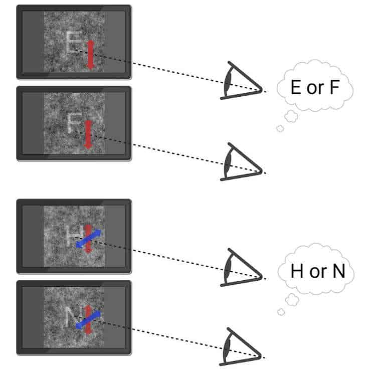
The researchers used sensitive equipment in Dr. Rucci’s laboratory to record ocular drifts in six volunteers who were asked to identify which of two letters (H vs. N, or E vs. F) was shown to them on a background of random visual noise. Based on computational modelling, the scientists predicted that optimal eye movements for letter discrimination would cross the key elements distinguishing the letters at right angles.
As a result, they hypothesized that, if such control existed, it would tend to direct ocular drift in both vertical and oblique (lower left to upper right) directions for the H vs. N discrimination, as opposed to more strictly vertical movements for the E vs. F discrimination.
Sensory and Motor Interrelationship
They discovered that the subjects’ eye movements did indeed tend to follow these patterns – even in the 20% of trials in which the subjects were shown only noise – despite expecting to see a letter. The latter result demonstrated that ocular drift cognitive control could be driven solely by specific prior knowledge of the visual task, independent of any incoming visual information.
“These results underscore the interrelationship between the sensory and the motor parts of vision — one really can’t view them separately,”
said Dr. Victor.
He noted that the direction of fine eye movements is believed to originate from neurons in the brainstem, whereas task-related knowledge resides in the cortex of the upper brain, implying a non-conscious connection between the two.
“The subjects are aware of the tasks they have to do, yet they don’t know that their eyes are executing these tiny movements, even when you tell them,”
Dr. Victor said.
He added that studies of this pathway could lead to a better understanding of not only the neuroscience of vision, but also visual disorders, which have traditionally been viewed as disorders of the retina or sensory processing in the brain.
Reference:
- Yen-Chu Lin et al, Cognitive influences on fixational eye movements, Current Biology (2023). DOI: 10.1016/j.cub.2023.03.026
Last Updated on September 20, 2023