Summary: A study led by researchers at the Broad Institute of MIT and Harvard has analyzed rare brain tissue samples from 52 living patients with varying degrees of Alzheimer’s-related changes in the brain, including 17 individuals later clinically diagnosed with the disease. The team identified a suite of changes in cells unique to the early stages of Alzheimer’s, including some not seen before in animal studies. They discovered a brief hyperactive state in a specific group of neurons that was associated with their death in later stages of the disease, and increased inflammatory processes in immune cells called microglia as the disease progressed.
The vast majority of Alzheimer’s disease research on human brain tissue has examined postmortem samples, making it challenging for scientists to identify the earliest events in the brain that may have triggered the accumulation of plaques and the death of neurons. Understanding the molecular changes that occur in neurons, glia, and other brain cells around plaques during the early stages of the disease could aid scientists in developing treatments that are most effective when administered early.
Now, in a study published in Cell, a team led by researchers from the Broad Institute of MIT and Harvard analyzed a collection of rare brain tissue samples from 52 living patients with varying degrees of other Alzheimer’s-related brain changes, including 17 individuals who were subsequently clinically diagnosed with the disease. Scientists identified a series of cell changes unique to the early phases of Alzheimer’s disease, including some not previously observed in animal research.
The researchers discovered a brief hyperactive state in a specific group of neurons that was associated with their death later in the disease, as well as increased inflammatory processes in immune cells called microglia as the disease progressed.
Neurons and Amyloids
The data collected by the researchers supported the hypothesis that neurons produce the plaque-forming protein amyloid beta. In addition, they discovered for the first time that oligodendrocytes, which generate insulating sheaths around the brain’s nerve fibers, may also contribute to plaque formation. An improved understanding of how these cells spur the growth of plaques could one day help researchers identify new targets for Alzheimer’s drugs.
The study is the result of close collaboration with Ville Leinonen, a neurosurgeon and professor at the University of Eastern Finland who has spent more than a decade collecting and studying brain tissue samples from patients who underwent routine surgeries for other neurological conditions and agreed to provide a small amount of brain tissue and other samples for research.
“This was just a really rich opportunity to peer into the actual workings of cells with minimal artifacts and see what they’re doing in the context of amyloid,”
said Evan Macosko, senior author on the study, an institute member at the Broad, and associate professor and attending psychiatrist at Massachusetts General Hospital.
Beth Stevens, an institute member at the Broad, an associate professor at Harvard Medical School, and a research associate in neurobiology at Boston Children’s Hospital, was a co-author on the study.
“This was a really great synergy of computational, wet lab, and clinical work. It took a decade of neurosurgeries, patient involvement, thoughtful analyses, and really useful experiments. We couldn’t have done this study if any one of those things hadn’t happened,”
said Tushar Kamath, co-first author on the study and an MD/Ph.D. student in Macosko’s lab when the study began.
Vahid Gazestani, a research scientist in Macosko’s lab when the study began and now a senior computational scientist at Johnson & Johnson, was the other co-first author of the study.
Postmortem Challenges
Cells — particularly neurons — change rapidly after losing their supply of oxygen postmortem, potentially making it difficult for scientists to accurately study how they work when just looking at postmortem samples. At a neuroscience meeting five years ago, Macosko was discussing these limitations with other researchers when a colleague suggested he speak with Leinonen.
Leinonen was studying early Alzheimer’s disease and normal pressure hydrocephalus (NPH), a neurological disorder characterized by excess fluid around the brain. He had a collection of brain tissue samples obtained from NPH patients during routine surgeries to reduce excess brain fluid.
He’d collected other samples, such as cerebrospinal fluid from the same patients, and followed the cohort over time, recording clinical data such as whether or not patients developed Alzheimer’s.
Macosko knew samples from these living patients presented a rare chance to observe cells exposed to the initial stages of Alzheimer’s pathology.
Single-nucleus Profiling
In the subsequent years, his team analyzed brain tissue using single-nucleus RNA sequencing, which maps gene expression in individual cell nuclei. By comparing this data with Leinonen’s clinical notes and integrating it with previous single-cell postmortem and mouse studies, the researchers were able to identify important changes in various cell types during the disease’s early stages.
“These samples gave us a high-quality anchor to reliably identify cell types in all the other datasets. This whole integrative analysis was feasible because of the quality and the depth of the dataset that we had,”
said Gazestani. Researchers have long assumed that neurons produce the amyloid protein, but this has been difficult to prove in human tissue.
In the new study, Macosko’s team found that neurons showed gene expression signatures associated with amyloid production. For the first time, they also observed the same signature in oligodendrocytes.
“This is exciting because there are lots of ways you can make amyloid accumulate in the brain in mouse studies, but now we’ve seen what’s actually happening in a human,”
Macosko said.
Upper Layer Hyperactivity
In addition, the researchers observed a hyperactive population of neurons in the brain’s upper layer. This group of neurons dies early in the disease, and Macosko believes that future research will demonstrate that this hyperactivity leads to a more extensive neuronal loss in patients.
Finally, the researchers identified microglia — cells that help clear the amyloid-beta peptide from the brain—that were functioning in two different kinds of activated states. Some of these states have not been detected previously in microglia in animal models, though the researchers recently confirmed several of these diverse states in induced pluripotent stem cells.
In the present study, the team found cells in one of these states in tissue from both patients with Alzheimer’s and those with Parkinson’s — a finding that could provide clues about similarities between the conditions.
In the future, Macosko and Stevens intend to identify proteins that correlate with these cell states in pairs of blood and cerebrospinal fluid samples, which could serve as disease progression markers. In addition, they anticipate that other researchers will use their method to analyze datasets from various sample types and sequencing techniques jointly.
Reference:
- Vahid Gazestani; Tushar Kamath; Naeem M. Nadaf; et al. Early Alzheimer’s disease pathology in human cortex involves transient cell states, Cell 0092-8674, Vol: 186, Issue: 20, Page: 4438-4453.e23
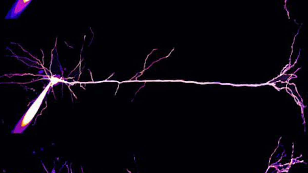
Image: pyramidal neuron in mouse visual cortex, credit Nguyen Tan Tin CC-BY