Researchers have developed a new brain imaging tool that can detect mild traumatic brain injuries (mTBIs) even when conventional imaging techniques such as magnetic resonance imaging (MRI) show no structural abnormalities. The method includes injecting gadolinium, a common MRI contrast agent, into hydrogel-based micropatches connected to immune cells known as macrophages.
mTBIs generate inflammation in the brain, which sends out signals that recruit macrophages. When these cells are mixed with the gadolinium contrast agent, MRI can find brain inflammation and more accurately identify mTBI patients, which leads to better treatment for patients.
“Seventy to ninety percent of reported TBI cases are categorized as ‘mild,’ yet as many as ninety percent of mTBI cases go undiagnosed, even though their effects can last for years and they are known to increase the risk of a host of neurological disorders including depression, dementia, and Parkinson’s disease. Our cell-based imaging approach exploits immune cells’ innate ability to travel into the brain in response to inflammation, enabling us to identify mTBIs that standard MRI imaging would miss,”
said senior author Samir Mitragotri, Ph.D. Mitragotri, in whose lab the research was conducted, is a Core Faculty member of the Wyss Institute at Harvard University and the Hiller Professor of Bioengineering and Hansjörg Wyss Professor of Biologically Inspired Engineering at Harvard’s John A. Paulson School of Engineering and Applied Sciences (SEAS).
mTBI Diagnosis Shortfall
Most of us know someone who has suffered a concussion (also known as a mTBI), and sometimes more than one. However, the vast majority of persons who suffer from mild traumatic brain injury are never properly diagnosed.
Without that diagnosis, patients may aggravate their injuries by returning to normal activity before they are fully recovered, causing severe damage. According to certain research, multiple mTBIs can lead to chronic traumatic encephalopathy (CTE), a neurodegenerative illness that has been reported to affect more than 90% of professional American football players.
Members of the Mitragotri lab made the decision to use their knowledge of immune cells to develop a better diagnostic tool because it is believed that “invisible” brain inflammation is what causes the effects of mTBI.
“Our previous projects have focused on controlling the behavior of immune cells or using them to deliver drugs to a specific tissue. We wanted to exploit another innate ability of immune cells—homing to sites of inflammation in the body—to carry imaging agents into the brain, where they can provide a visible detection signal for mTBI,”
said first author Lily Li-Wen Wang, Ph.D., a former Research Fellow in the Mitragotri Lab at the Wyss Institute and SEAS.
Hydrogel Backpack
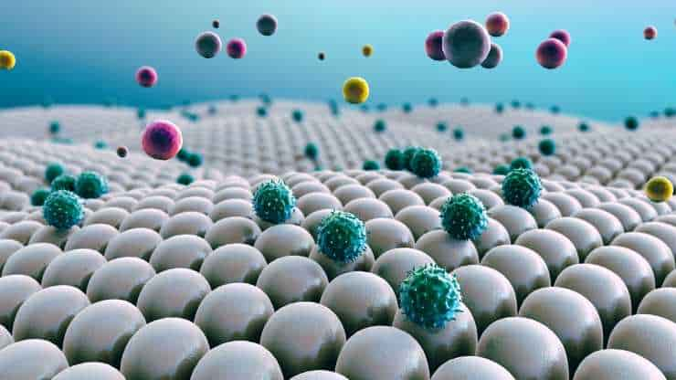
The scientists planned to attach gadolinium molecules to macrophages, a type of white blood cell known to invade the brain in response to inflammation, using the cellular backpack method. But they quickly ran into a problem: gadolinium must interact with water in order to operate as a contrast agent for MRI imaging.
Their original backpack microparticles are made from a polymer called PLGA, which is hydrophobic (repels water). So Wang and her co-authors began working on a novel backpack comprised of a hydrogel material that could be mass-produced in the lab.
Finally, they developed a new hydrogel backpack that could create a strong gadolinium-mediated MRI signal, stick to macrophages from both mice and pigs, and keep their cargo for a long time in the lab.
They named their new microparticles M-GLAMs, short for “macrophage-hitchhiking Gd(III)-Loaded Anisotropic Micropatches.” Now, it was time to test them in a more realistic setting, for which they partnered with researchers and clinicians at Boston Children’s Hospital.
M-GLAM Testing
They started by injecting mouse M-GLAMs macrophages into mice to test if they could spot them in vivo. They were particularly interested in seeing if they accumulated in the kidney, because existing gadolinium-based contrast agents, such as Gadavist, can pose health problems to patients with kidney illness.
Their M-GLAMs did not accumulate in the mice’s kidneys, but persisted in their bodies for over 24 hours with no negative side effects. In contrast, mice injected with Gadavist showed substantial accumulation of the contrast agent in their kidneys within 15 minutes of injection, and the substance was fully cleared from their bodies within 24 hours.
The researchers next put porcine M-GLAMs to the test in a pig model of mTBI. The M-GLAMs were infused into the animals’ blood two days after a simulated mTBI, and the concentration of gadolinium in the brain was measured by MRI. They concentrated on the choroid plexus, a tiny region regarded as a main route of immune cells into the brain.
Increased Gadolinium Intensity
It was found that pigs given M-GLAMs had a significant increase in the amount of gadolinium in the choroid plexus, but pigs given Gadavist did not. This was true even though there were more inflammatory macrophages in the brains of both groups. Following administration of the medicines, the animals showed no harm in any of their main organs.
“Another important aspect of our M-GLAMs is that we are able to achieve better imaging at a much lower dose of gadolinium than current contrast agents—500-1000-fold lower in the case of Gadavist,” said Wang. “This could allow the use of MRI for patients who are currently unable to tolerate existing contrast agents, including those who have existing kidney problems.”
While M-GLAMs can suggest the presence of inflammation in the brain due to the high concentration of macrophages entering it through the choroid plexus, the scientists point out that their technique cannot pinpoint the precise location of lesions or inflammatory reactions in brain tissue. However, when combined with new treatment modalities developed in another recent publication, M-GLAMS could provide a more quick and effective way to identify and control inflammation in mTBI patients, minimizing damage and speeding recovery.
Immune Cell Ingenuity
The researchers have applied for a patent for their technique and intend to bring it to market in the near future. They are currently looking into cooperation with biotech and pharmaceutical businesses to help them get to clinical trials faster.
“This work demonstrates just how much potential is waiting to be unlocked within the human body for a variety of functions: monitoring health, diagnosing problems, treating diseases, and preventing their recurrence. I’m impressed with this team’s ingenuity in leveraging immune cells to improve medical imaging, and hope to see it in clinicians’ hands soon,”
said Wyss Founding Director Donald Ingber, M.D., Ph.D. Ingber is also the Judah Folkman Professor of Vascular Biology at Harvard Medical School and Boston Children’s Hospital, and the Hansjörg Wyss Professor of Bioinspired Engineering at SEAS.
Abstract
The choroid plexus (ChP) of the brain plays a central role in orchestrating the recruitment of peripheral leukocytes into the central nervous system (CNS) through the blood-cerebrospinal fluid (BCSF) barrier in pathological conditions, thus offering a unique niche to diagnose CNS disorders. We explored whether magnetic resonance imaging of the ChP could be optimized for mild traumatic brain injury (mTBI). mTBI induces subtle, yet influential, changes in the brain and is currently severely underdiagnosed. We hypothesized that mTBI induces sufficient alterations in the ChP to cause infiltration of circulating leukocytes through the BCSF barrier and developed macrophage-adhering gadolinium [Gd(III)]–loaded anisotropic micropatches (GLAMs), specifically designed to image infiltrating immune cells. GLAMs are hydrogel-based discoidal microparticles that adhere to macrophages without phagocytosis. We present a fabrication process to prepare GLAMs at scale and demonstrate their loading with Gd(III) at high relaxivities, a key indicator of their effectiveness in enhancing image contrast and clarity in medical imaging. In vitro experiments with primary murine and porcine macrophages demonstrated that GLAMs adhere to macrophages also under shear stress and did not affect macrophage viability or functions. Studies in a porcine mTBI model confirmed that intravenously administered macrophage-adhering GLAMs provide a differential signal in the ChP and lateral ventricles at Gd(III) doses 500- to 1000-fold lower than those used in the current clinical standard Gadavist. Under the same mTBI conditions, Gadavist did not offer a differential signal at clinically used doses. Our results suggest that macrophage-adhering GLAMs could facilitate mTBI diagnosis.
Reference:
- Lily Li-Wen Wang et al. Preclinical characterization of macrophage-adhering gadolinium micropatches for MRI contrast after traumatic brain injury in pigs. Sci. Transl. Med. 16, eadk5413 (2024) DOI:10.1126/scitranslmed.adk5413