Researchers at the Max Planck Institute for Brain Research and Goethe University Frankfurt have uncovered a neural pathway in the rodent brain that may play a key role in processing spatial perspectives and help the animal to detect boundaries to avoid bumping into things.
Animals use landmarks in the environment as a reference point to identify the self’s position and navigate their surroundings. In rodents, this ability is supported by very specialized types of neurons, including place cells and grid cells, that fire only when the animal is at a precise location in the environment, even in an open arena,
says Hiroshi Ito, senior author of the study.1
Retrosplenial Cortex
You perceive the world relative to your own body from a self-centered perspective. But your brain is able to translate this information into a world-centered, cognitive map of the environment, guiding us independent of where you look or the direction you face.
Neuroscientists know that the retrosplenial cortex is an important brain area for processing this landmark information. However, the exact function of individual neurons in this brain region is still unknown.
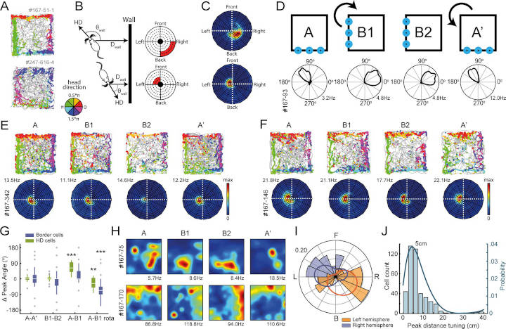
By recording from the retrosplenial cortex in the rat, we discovered a new type of neuron that signals the location of the room’s boundaries such as walls from the animal’s perspective. Border cells fire with high precision. In this case, only when the boundary is at a particular distance and direction away from the animal,
says lead author Joeri van Wijngaarden.
But how do border cells know when to be active? Do they use direct sensory cues, such as vision or their whiskers, to detect the walls? To investigate these questions, the researchers manipulated the sensory experience of the animal.
To our big surprise, we saw no difference when the rat explored the maze in complete darkness. The cells kept firing as they did before,
says van Wijngaarden.
Border Cells And Spatial Cells
The scientists wanted to then investigate how border cells interact with spatial cells in a connected brain region, the entorhinal cortex, that is crucial for spatial processing and forming an internal map of the environment. They approached the problem by recording activity from spatial cells in the entorhinal cortex while silencing border cells in the retrosplenial cortex with a drug.
At first, we saw no effect. However, when we switched gears and instead silenced spatial cells in the entorhinal cortex, we suddenly noticed a disruption of border cell activity in the retrosplenial cortex. This was a big surprise as it suggests that border cells capture landmark information without the need to sense it directly. Instead, they rely on spatial information from other brain areas to calculate their position,
explains van Wijngaarden. What struck him above all was the close correlation between the activity of these neurons and the animal’s following motion.
When the rat approaches a wall to the left, border cells in the right hemisphere are activated, just before the animal turns right. Conversely, border cells in the left hemisphere are active just before left turns, as to avoid collision,
he says.
The findings bring a new perspective to the field, providing the first glimpse into how the brain’s internal map can be used to guide our moves during navigation.
- Joeri BG van Wijngaarden et al.
Entorhinal-retrosplenial circuits for allocentric-egocentric transformation of boundary coding.
eLife 2020;9:e59816 DOI: 10.7554/eLife.59816 ↩︎
Last Updated on October 15, 2022