Since their discovery in the 1990s, the brain’s head-direction cells have been dubbed its “internal compass.” These cells are assumed to be vital for spatial orientation and navigation since they are triggered when an animal’s or human’s head points in a specific direction.
A team of neuroscientists from the University of Tübingen has now revealed that head-direction cells in mice accomplish much more. They may be involved in the transmission of sensory and emotional information used to construct memories of experiences, a process known as episodic memory.
“The hippocampus is a kind of neural curator that integrates the information. During an experience, a memory trace is created in the hippocampus for that episode in our lives,”
Said Professor Andrea Burgalossi from the Institute of Neurobiology and the Werner Reichardt Center for Integrative Neuroscience (CIN), who led the research team.
Enter the Anterior Thalamus
In the external world of human experience, the senses together contribute to the formation of memories.
The visual stimulus of a beautiful landscape, the echo of a laugh, the warmth of a hug — all of these sensory stimuli are brought together in the hippocampus, an area of the brain. This processing is essential for converting transitory sensory inputs into long-term memories.
The research team concentrated on one of the hippocampus’s main input structures in the brain, the anterior thalamus, to better understand where sensory information enters the hippocampus.
“We have known for decades that this area is crucial for episodic memory. Patients with damage to this region of the brain suffer from memory loss,”
said Dr. Patricia Preston-Ferrer, one of the lead authors of the study.
Sensory Stimuli Activity
Scientists discovered head-direction cells in rodents’ anterior thalamus when they first recorded the activity of nerve cells there in the 1990s.
“Previously, it was assumed that these only encoded the animal’s heading direction in its environment,” says Preston-Ferrer. “But now our latest experiments show that this idea provides an incomplete picture.”
When the Tübingen research team recorded the electrical activity in the mouse brain, they discovered that when the mouse was exposed to sensory stimuli, the head-direction cells in the thalamus became active.
“In the case of a sound being played, as well as in the case of a tactile whisker on the mouse’s snout being touched, only the head-direction cells were activated specifically and reliably and with a remarkably short delay. We were surprised, as it had been assumed for decades that these neurons were unresponsive to sensory stimuli,”
said CIN researcher and co-author of the study Giuseppe Balsamo.
Episodic Memory Involvement
The experiments revealed that in the anterior thalamus, only the head-direction cells responded to sensory stimuli.
“This tells us that head-direction cells must have a special function,”
said co-author Dr. Eduardo Blanco-Hernandez.
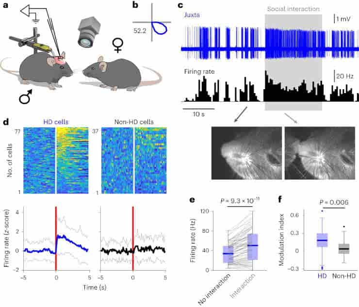
Their role must go beyond that of an internal compass. The head-direction cells also increased their activity in response to aroused circumstances, such as meeting another mouse.
“It is known that close attention and emotions have a great influence on the formation of memories and their quality. In such situations, we remember much more vividly than in an uninvolved, passive state,”
said Blanco-Hernandez.
Overall, the new findings suggest that head-direction cells in the thalamus may serve as a key gateway for sensory, attention, and arousal information entering the episodic memory system.
“To understand how a memory trace is formed, we need to know the pathways and nerve cells involved that transmit basic information to the hippocampus. Based on our work, we believe the inner compass represents a key node in this process,”
said Burgalossi
Further research will be required to determine whether this node can be influenced, for example, for therapeutic purposes in order to better form and retrieve memories.
Abstract
Head-direction (HD) neurons are thought to exclusively encode directional heading. In awake mice, we found that sensory stimuli evoked robust short-latency responses in thalamic HD cells, but not in non-HD neurons. The activity of HD cells, but not that of non-HD neurons, was tightly correlated to brain-state fluctuations and dynamically modulated during social interactions. These data point to a new role for the thalamic compass in relaying sensory and behavioral-state information.
Reference:
- Blanco-Hernández, E., Balsamo, G., Preston-Ferrer, P. et al. Sensory and behavioral modulation of thalamic head-direction cells. Nat Neurosci (2024). Doi: 10.1038/s41593-023-01506-1
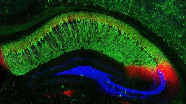
Top image: Mouse hippocampus cross section. The large, crescent-shaped area in green is hippocampal zone CA1. Its highly specialized neurons, called place cells, serve as the brain’s GPS system to track location. Credit: NIH/Raunak Basu, University of Utah, Salt Lake City