A set of galanin producing nerve cells, located in a region of the hypothalamus called the ventrolateral preoptic nucleus (VLPO), are essential to normal sleep, research from Beth Israel Deaconess Medical Center has found.
The study build on work from two decades ago, when Clifford B. Saper, MD/Ph.D., Chairman of the Department of Neurology at Beth Israel Deaconess Medical Center (BIDMC), and colleagues discovered a set of nerve cells they thought might be the switch that turns the brain off, allowing it to sleep.
“Our paper is the first test of what happens when you activate the VLPO neurons. The findings support our original observation that the VLPO cells are essential to normal sleep,”
said Saper, who is also James Jackson Putnam Professor of Neurology and Neuroscience at Harvard Medical School.
Contradiction Resolved
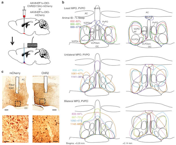
Working with genetically engineered mice, Saper’s team artificially activated the VLPO neurons using several different tools.
In one set of experiments, the scientists activated the neuron cells using a laser light beam to make them fire, a process called optogenetics. In another test, the team used a chemical to selectively activate the VLPO neurons. In both cases, activating these cells profoundly drove sleep.
The results confirmed Saper and colleagues’ earlier findings that these neurons are active during sleep and that damage to them causes insomnia — as seen in Saper’s subsequent work with laboratory animals and, in 2014, in older people who have lost cells of the VLPO as part of the natural aging process.
Based on that previous body of work, it came as a surprise when another team of researchers reported just the opposite. In a 2017 publication, experiments stimulating the VLPO neurons woke laboratory animals up. In their current paper, Saper’s team cleared up the seeming contradiction.
Preoptic Area
The preoptic area (POA) is necessary for sleep, but the fundamental POA circuits have remained elusive, and the neurochemical identity of sleep-promoting neurons has remained controversial.
“We found that when the VLPO cells are stimulated one to four times per second, they fire each time they are stimulated, resulting in sleep,” Saper said. “But if you stimulate them faster than that, they begin to fail to fire and eventually stop firing altogether. We learned our colleagues in the other lab were stimulating the cells 10 times per second, which was actually shutting them off.”
Additionally, Saper’s team also found that activating the VLPO cells caused a fall in body temperature.
Scientists already knew that warm temperatures activate VLPO cells, and that body temperature dips slightly during sleep, when the VLPO neurons are firing.
“We thought that this is why people need to curl up under a warm blanket to get to sleep,”
Saper added.
However, with continued activation, body temperature in the mice fell by as much as five or six degrees Celsius. Saper’s team proposed that excessive firing of these same neurons may be responsible for the prolonged sleep and decline in body temperature in animals that hibernate.
In follow-up, Saper’s team is already looking at the relationship between sleep and body temperature in ongoing studies.
Daniel Kroeger, Gianna Absi, Celia Gagliardi, Sathyajit S. Bandaru, Joseph C. Madara, Loris L. Ferrari, Elda Arrigoni, Heike Münzberg, Thomas E. Scammell, Clifford B. Saper & Ramalingam Vetrivelan
Galanin neurons in the ventrolateral preoptic area promote sleep and heat loss in mice
Nature Communications volume 9, Article number: 4129 (2018_
Related Posts:
- How Neurons Adjust Excitation-Inhibition Balance Unaided
- Brains Work Via Their Genes Just As Much As Their Neurons
- Do Mental Map Neurons Play A Wider Cognitive Role?
Last Updated on December 7, 2022