A very high resolution map of the white-grey matter border across the entire living brain has been created by a multidisciplinary team led by Nikolaus Weiskopf from the Max Planck Institute for Human Cognitive and Brain Sciences.
Current neuroscience thinks of the brain as composed of two tissue types. Billions of neurons make up the gray matter, forming a thin layer on the brain’s surface. These neuronal cells are interlinked in a network by hundreds of millions of white matter connections, running in bundles, deeper in the brain.
Until recently, little was known about the interface between the white and gray matter — known as superficial white matter — because methods were lacking to study it in living human brains. Yet, previous investigations had suggested the region to be involved in serious conditions like epilepsy, Alzheimer’s disease and autism.
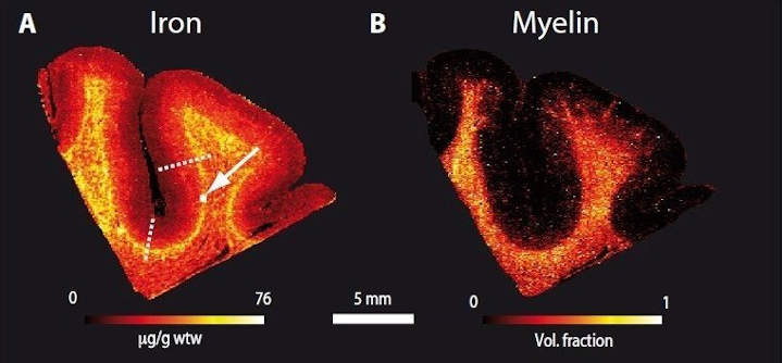
“We demonstrated that the superficial white matter contains a lot of iron. It is known that iron is necessary for the process of myelination,”
says Dr. Evgeniya Kirilina, first author of the study.1
Frontal Cortex Myelination
Myelin, which gives white matter it’s white coloring, is the fatty coating of nerve cell axons that speeds up transmission of information through the brain. The myelination process can occur throughout the lifespan but is predominant during development.
In fact, the largest concentration of iron the researchers found was in the superficial white matter in regions of the frontal cortex, which happens to be the slowest developing structure in the human brain. In humans, our frontal cortex is not fully myelinated until our fourth decade of life.
Key to the new method is Magnetic Resonance Imaging (MRI), but at very high field strength.
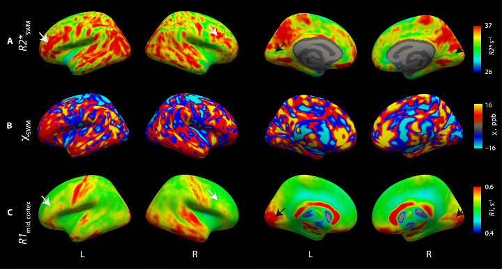
While typical clinical MRI scanners work at 1.5 or 3 Tesla, in terms of the strength of the magnetic field, the Max Planck Institute for Human Cognitive and Brain Sciences houses a powerful 7 Tesla scanner. In combination with an advanced biophysical model, powerful MRI enabled the researchers to create very high resolution maps of the white-gray matter border across the entire living brain.
The accuracy of their submillimetre maps was assessed against classic and advanced histological methods involving physical dissection and analysis of post-mortem brains.
The new method promises many further insights into the organization of the interface between white and gray matter.
We hope the method can be used to increase our understanding of brain development as well as pathological conditions involving the superficial white matter,
Evgeniya Kirilina adds.
The work was supported by the German Research Foundation, the Alzheimer-Forschung-Inititiative, the European Union’s Seventh Framework Programme, and the Swiss State Secretariat for Education, Research and Innovation (SERI).
- Evgeniya Kirilina, Saskia Helbling, Markus Morawski, Kerrin Pine, Katja Reimann, Steffen Jankuhn, Juliane Dinse, Andreas Deistung, Jürgen R. Reichenbach, Robert Trampel, Stefan Geyer, Larissa Müller, Norbert Jakubowski, Thomas Arendt, Pierre-Louis Bazin, Nikolaus Weiskopf; Superficial white matter imaging: Contrast mechanisms and whole-brain in vivo mapping. Science Advances 07 Oct 2020: Vol. 6, no. 41, eaaz9281 DOI: 10.1126/sciadv.aaz9281 ↩︎
Last Updated on October 4, 2022