Acute stress is associated with increased myelination of axons in areas of the brain associated with memory and emotions, recent research shows.
The study1, from scientists at the University of California, Berkeley, and UC San Francisco (UCSF), represents a potential explanation for why some people are quick to recover from, and others vulnerable to traumatic stress, and for the varied symptoms – avoidance behavior, anxiety and fear, for example – triggered by the memory of such stress.
If, as the researchers suspect, extreme trauma causes the increased myelination, the findings could lead to treatments – drugs or behavioral interventions – that prevent or reverse the myelin production and lessen the aftereffects of extreme trauma.
Increased Gray Matter Myelin
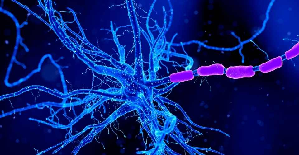
Myelin wraps around the axons of neurons to facilitate long-distance transmission of signals and, thus, communication between distant areas of the brain. The inner regions of the brain look white; in fact, they are referred to as white matter, because of the myelin encasing the many large bundles of axons there.
But this study uncovered increased myelination of axons in so-called gray matter, where most of the cell bodies of neurons reside and most of the wiring is less insulated with myelin. The extra myelination was found primarily in brain areas associated with memory.
Researchers from San Francisco Veterans Affairs Medical Center conducted brain MRI scans of 38 veterans – half with PTSD, half without – and found an increase in myelination in the gray matter of those with PTSD compared to that seen in the brains of those not suffering from PTSD. Concurrently, colleagues at UC Berkeley, discovered a comparable boost in myelination in the gray matter of adult rats subjected to an acute stressful event.
While not all rats showed long-term effects from the stress, just as not all traumatized veterans develop PTSD, those that did had increased myelination in specific areas of the brain associated with particular symptoms of stress that was identical to what UCSF physicians found in veterans with PTSD.
Directions In Treatment
Both veterans with PTSD and stressed rats that exhibited avoidance behavior had increased myelination in the hippocampus, often thought of as the seat of memory. Those exhibiting a fear response had increased myelination in the amygdala, which plays a key role in our response to strong emotions, such as fear or pleasure. Those suffering from anxiety had increased myelination in the dentate gyrus, a region critical to learning and memory.
The combination of these studies in rats with our population of veterans with post traumatic stress disorders is, to me, really exciting. At least it’s another mechanism to think about as we develop new treatments. If we see enduring ability to shape myelin content in an adult brain, maybe treatments will help reverse this. That’s where we want to go next with this,
said senior author Dr. Thomas Neylan.
The study suggests that the specific symptoms are related to which areas of the brain are being newly myelinated.
There’s a lot of heterogeneity across different people with PTSD; it’s not one size fits all. Every PTSD patient generally has a mix of different symptoms. Some people are very avoidant. Some people are very hyperreactive. The idea is that if you can show that these different symptom clusters have different neural circuitry, it might actually lead us closer to subtyping people in a way that we could be more targeted in our treatment,
said Neylan, professor-in-residence in psychiatry at the UC San Francisco Weill Institute for Neurosciences.
Hyperresponsive Connections
The researchers show that stress produces more of the brain’s glial cells, called oligodendrocytes, which wrap around the axons of neurons and make the myelin. The increased myelin produced by these new oligodendrocytes could affect the speed of connections between neurons2, making some connections hyperresponsive.
In the gray matter of your cortex, most of the dendrites and axons, the projections that come out of the neurons that help establish communications with other neurons, can form thousands of connections, and most of them are unmyelinated. But if experience leads you to start to lay down myelin to strengthen certain connections, let’s say your ability to respond quickly to a fearful stimulus, you can speed up that circuit, but you lose the kind of broader adaptive flexibility that you normally would have with mostly unmyelinated axons and dendrites. People with PTSD become almost like a one-note musician – they really know how to respond to fear. But that enhanced, quick response to fear may diminish their adaptive flexibility for non-fear-type behavior,
Neylan said.
The researchers are collaborating on further studies, including looking for increased myelin in the brains of PTSD patients who have died, improving fMRI imaging of myelin in the brain, investigating the effects of chronic stress on the brain connections of rats, and using new high-resolution imaging to study the myelin deposition in gray matter.
- Kimberly L. P. Long, Linda L. Chao, Yurika Kazama, Anjile An, Kelsey Y. Hu, Lior Peretz, Dyana C. Y. Muller, Vivian D. Roan, Rhea Misra, Claire E. Toth, Jocelyn M. Breton, William Casazza, Sara Mostafavi, Bertrand R. Huber, Steven H. Woodward, Thomas C. Neylan, Daniela Kaufer. Regional gray matter oligodendrocyte- and myelin-related measures are associated with differential susceptibility to stress-induced behavior in rats and humans Translational Psychiatry, 2021; 11, 631 ↩︎
- Liu J, Dietz K, Hodes GE, Russo SJ, Casaccia P. Widespread transcriptional alternations in oligodendrocytes in the adult mouse brain following chronic stress. Dev Neurobiol. 2018;78:152–62. ↩︎
Last Updated on October 26, 2022