The parts of the brain that support our ability to solve problems without prior experience — also known as fluid intelligence — have been mapped by a team led by University College London researchers.
Fluid intelligence could just be the defining characteristic of human cognition. It forecasts academic and professional achievement, social mobility, health, and longevity. It correlates with a variety of cognitive abilities, including memory.
It is believed that fluid intelligence is a crucial component of “active thinking” — a set of complex mental processes that includes abstraction, judgment, attention, strategy generation, and inhibition. All of these skills are applicable to daily tasks, from planning a dinner party to filing taxes.
Lesion-deficit Mapping
Despite its central role in human behaviour, the nature of fluid intelligence’s relationship with the brain and whether it is a single or a collection of cognitive abilities remains controversial.
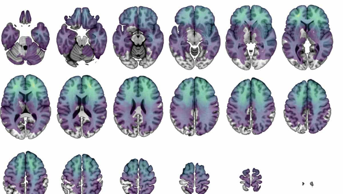
To determine which brain regions are necessary for a particular skill, researchers must examine patients whose brain regions are missing or damaged. These “lesion-deficit mapping” studies are challenging to conduct due to the difficulty in identifying and evaluating patients with focal brain injury.
As a result, functional imaging (fMRI) techniques have been primarily used in earlier studies; the results can be deceptive.
Raven Advanced Progressive Matrices
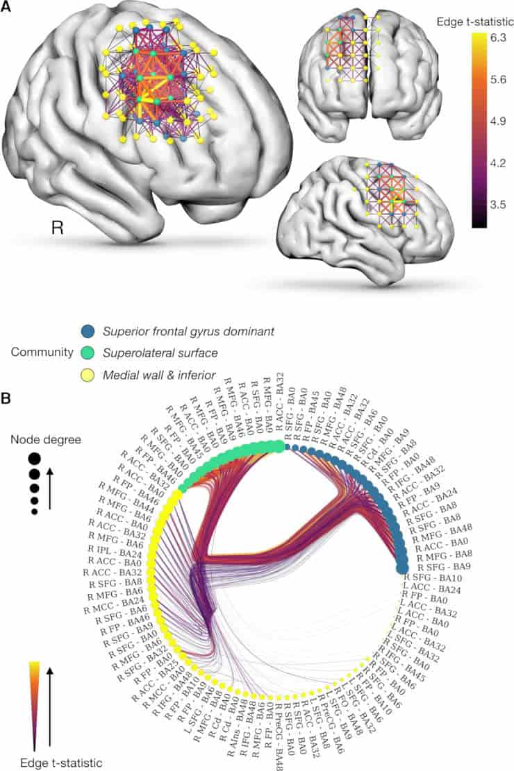
(A) Network-based statistics identify a significant network associated reduced adjusted APM scores (FWER-P < 0.0001).
(B) Radial graph of the community structure of the network inferred from a stochastic block model of its statistics shows that the network clusters into three discrete components encompassing the superolateral cortical surface, the medial (and inferior) wall and a superior frontal gyrus dominant cluster.
Credit: Lisa Cipolotti, James K Ruffle et al. Brain, awac304 CC-BY
Researchers from the National Hospital for Neurology and Neurosurgery at UCLH and the UCL Queen Square Institute of Neurology used the Raven Advanced Progressive Matrices (APM), the most well-known test of fluid intelligence, to examine 227 patients who had either experienced a brain tumour or a stroke to particular parts of the brain.
There are increasing difficulty multiple-choice visual pattern questions on the test. Each issue asks you to choose the final piece from various options out of a pattern of incomplete geometric figures.
The researchers then devised a cutting-edge “lesion-deficit mapping” strategy to unravel the complex anatomical patterns of prevalent types of brain injury, like stroke. Their method looked at the connections between brain areas as a mathematical network.
The links between the areas show how likely they are to be affected together, either because of how the disease works or because they have similar cognitive abilities.
This allowed researchers to map the various parts of the brain and identify which patients performed worse on the fluid intelligence task in relation to their injuries. They were also able to disentangle the brain map of cognitive abilities from the patterns of damage.
Right Frontal Regions
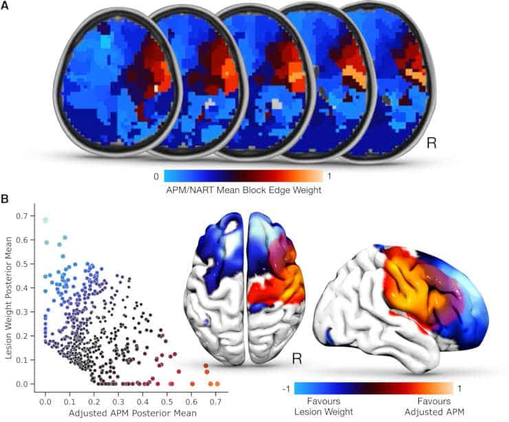
(B.) Scatterplot illustrating the relationship between posterior mean edge weights at each community block, for both the lesion weight (y-axis) and adjusted APM (x-axis), with brain reconstructions overlaying these findings.
Credit: Lisa Cipolotti, James K Ruffle et al. Brain, awac304 CC-BY
Instead of affecting a large number of brain regions distributed throughout the body, the researchers discovered that fluid intelligence impairment in performance was primarily limited to patients with right frontal lesions. Such damage is frequently observed in patients with a variety of neurological conditions, such as traumatic brain injury and dementia, in addition to brain tumours and stroke.
“Our findings indicate for the first time that the right frontal regions of the brain are critical to the high-level functions involved in fluid intelligence, such as problem solving and reasoning. This supports the use of APM in a clinical setting, as a way of assessing fluid intelligence and identifying right frontal lobe dysfunction,”
said lead author Professor Lisa Cipolotti.
The method used by the researchers, which combines novel lesion-deficit mapping with in-depth analysis of APM performance in a sizable patient sample, sheds light on the neural underpinnings of fluid intelligence. The relationship between the brain and cognition, which frequently determines how neurological disorders are treated, must receive more attention in lesion studies.
Reference:
Lisa Cipolotti, James K Ruffle, Joe Mole, Tianbo Xu, Harpreet Hyare, Tim Shallice, Edgar Chan, Parashkev Nachev. Graph lesion-deficit mapping of fluid intelligence. Brain, 2022;, awac304,