A novel imaging technique provides structural information about brain tissue that was previously difficult to access.
Diattenuation Imaging (DI), developed by scientists at Forschungszentrum Jülich and the University of Groningen, enables researchers to distinguish regions with many thin nerve fibres from regions with few thick nerve fibres. With existing imaging methods, these tissue types cannot easily be differentiated.
The DI method is based on 3-D polarized light imaging (3-D-PLI), a neuroimaging technique developed at Forschungszentrum Jülich, which reveals nerve fibre pathways with micrometre resolution. The technique is used, for instance, in the European Human Brain Project to investigate the 3-D fibre structures of the brain in unprecedented detail.
Polarized Light
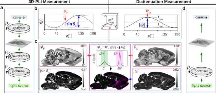
During a 3-D polarized light imaging measurement, histological brain sections are illuminated with polarized light. Depending on how the direction of oscillation (polarization) is oriented relative to the nerve fibres, the light is refracted to different degrees, allowing the computation of the spatial orientation of the nerve fibres.
This effect, called birefringence, is mainly caused by the myelin sheath, an insulating layer that surrounds many nerve fibres in the brain.
While 3-D-PLI measures the polarization-dependent refraction of light, a diattenuation measurement determines the polarization-dependent attenuation of light, i.e. how much the intensity of polarized light is reduced when passing through the brain section. The measurement is performed with the same apparatus as 3-D-PLI, whereby two filters are removed.
Diattenuation Imaging
The scientists discovered that diattenuation imaging — a combined measurement of diattenuation and 3-D-PLI — can distinguish between different brain regions. In some regions, the brain tissue is maximally transparent when the polarization of the light is oriented parallel to the nerve fibres.
In other regions, the tissue is maximally transparent when the polarization is oriented perpendicularly to the nerve fibres. How the tissue behaves depends, among other things, on the time after embedding the brain sections.
Using simulations on the Jülich a high-scaling supercomputer JUQUEEN, the researchers could show that the observed effects also depend on other tissue properties like the diameter of the fibres or the thickness of the myelin sheaths. This makes diattenuation imaging a valuable extension to 3-D-PLI, enabling a more precise investigation of brain tissue.
In the future, the DI method could be used to study neurodegenerative diseases like multiple sclerosis or multisystem atrophy (MSA), which go along with alterations of the myelin sheath. In addition, the technology helps to make pathological changes visible and to identify connected regions and tissue types, assisting the complex reconstruction of the brain.
The work received funding from the Helmholtz Association portfolio theme ‘Supercomputing and Modeling for the Human Brain’, from the European Union’s Horizon 2020 Research and Innovation Programme, and from the National Institutes of Health.
Reference:
- Miriam Menzel, Markus Axer, Katrin Amunts, Hans De Raedt & Kristel Michielsen. Diattenuation Imaging reveals different brain tissue properties. Scientific Reports volume 9, Article number: 1939 (2019) DOI: 10.1038/s41598-019-38506-w
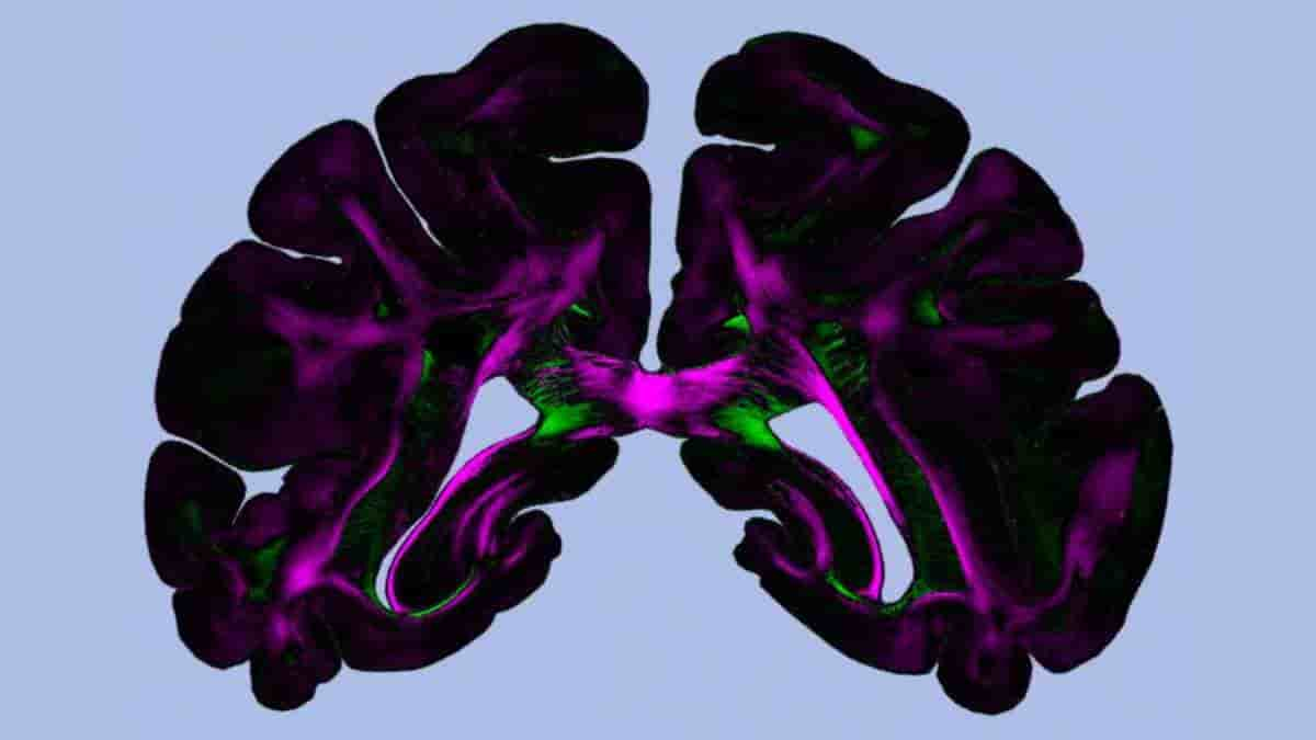
Top Image: The colors reveals the direction of polarization for which a maximum amount of light passes through the brain section. Regions for which this polarization direction runs parallel (or perpendicular) to the fiber direction are marked in green (or magenta). Credit: Miriam Menzel et al., Scientific Reports (2019), DOI:10.1038/s41598-019-38506-w (CC BY 4.0)
Last Updated on October 27, 2023