Four types of neurons in the peripheral auditory system, three of which are new to science, have been identified by researchers at Karolinska Institutet in Sweden. The analysis of these cells could lead to new therapies for various kinds of hearing disorders, such as tinnitus and age-related hearing loss.
When sound reaches the inner ear, it is converted into electrical signals that are relayed to the brain via the ear’s nerve cells in cochlea. Previously, most of these cells were considered to be of two types: type 1 and type 2 neurons, type 1 transmitting most of the auditory information.
This new study shows that the type 1 cells actually comprise three very different cell types, which tallies with earlier research showing variations in the electrical properties and sonic response of type 1 cells.
“We now know that there are three different routes into the central auditory system, instead of just one. This makes us better placed to understand the part played by the different neurons in hearing. We’ve also mapped out which genes are active in the individual cell types,”
says Francois Lallemend, research group leader at the Department of Neuroscience, Karolinska Institutet, who led the study.
Decoding Sonic Intensity
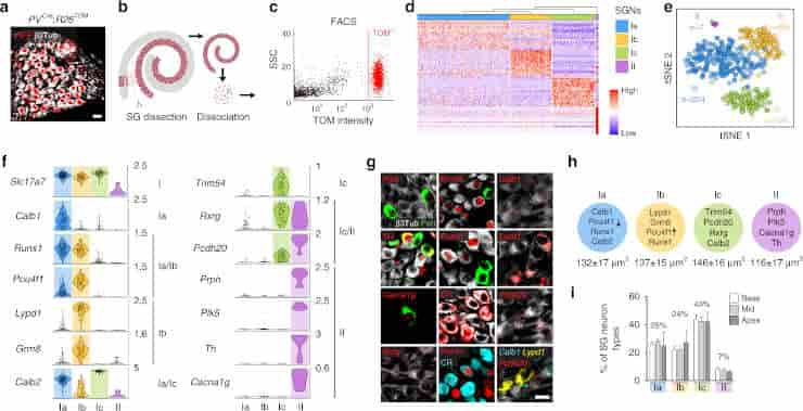
a) Genetic tracing of SG neurons (β3Tub+/RFP+) on P21 sections from PVcre;R26TOM mice.
(b) Sketch depicting dissection of SG from the organ of Corti and their dissociation.
(c) Fluorescence-side scatter plot of dissociated single cells showing isolation of Tom+ SG neurons through FAC sorting.
(d) Heat map showing single-cell expression of the top 20 differentially expressed genes in the four types of SG neurons, from combined data of P17, P21, and P33 neurons (SGNs, SG neurons). Dendritic tree shows the similarity between neuronal types.
(e) tSNE plot showing four distinct types of SG neurons.
(f) Violin plots showing the expression of marker genes in log-transformed scale among the four different populations of SG neurons.
(g) In vivo validation of the identified SG neuron types by immunohistochemical and fluorescent in situ hybridization using identified marker genes in P21 cochlea.
Type II neurons were identified by peripherin (Peri), Plk5, TH, and Cacna1g specifically. Ia neurons were identified by Calb1, Pou4f1, Runx1, and calretinin (CR). Ib neurons were identified by Lypd1, Runx1, and Pou4f1 and Ic neurons, by Rxrg, Pcdh20, and CR expression.
Note that co-localization on sections could never be observed for markers expressed in different populations of neurons in the scRNAseq data.
(h) Schematic representation of neuronal types with their key markers and their average soma size (in µm2) at P21.
(i) Proportion of SG neurons types along the tonotopic gradient (from base to apex) quantified by Runx1, Peri, and CR expression at P21 (n = 3 animals; Data are represented as mean ± SEM). Scale bars: 20 μm (a,g)
Credit: Charles Petitpre, et al. CC-BY
The team conducted their study on mice using the relatively new technique of single-cell RNA sequencing. The result is a catalogue of the genes expressed in the nerve cells, which can give scientists a solid foundation for better understanding the auditory system as well as for devising new therapies and drugs.
“Our study can open the way for the development of genetic tools that can be used for new treatments for different kinds of hearing disorders, such as tinnitus,” says Dr Lallemend. “Our mapping can also give rise to different ways of influencing the function of individual nerve cells in the body.”
The study shows that these three neuron types probably play a part in the decoding of sonic intensity (i.e. volume), a function that is crucial during conversations in a loud environment, which rely on the ability to filter out the background noise.
This property is also important in different forms of hearing disorders, such as tinnitus or hyperacusis (oversensitivity to sound).
Neuron Types In The Cochlea
Sound is transduced in the cochlea by hair cells (HCs), which then convey signals to the central nervous system through chemical synapses on the spiral ganglion (SG) neuron’s dendrites.
Most SG neurons are type I neurons; they are myelinated, contact the inner hair cells (IHCs) and are the principal carrier of the auditory signal. The other minority population (5%) of neurons are called type II neurons; they are unmyelinated and innervate the outer hair cells (OHCs), which modulate the output of the organ of Corti.
However, the cellular basis of processing and routing these auditory qualities at the periphery is still poorly understood.
“Once we know which neurons cause hyperacusis we’ll be able to start investigating new therapies to protect or repair them,” explains Dr Lallemend. “The next step is to show what effect these individual nerve cells have on the auditory system, which can lead to the development of better auditory aids such as cochlear implants.”
The researchers have also shown through comparative studies on adult mice that these different types of neurons are already present at birth.
The work was supported by grants from the Swedish Medical Research Council, the Knut and Alice Wallenbergs Foundation, the Swedish Brain Foundation, Karolinska Institutet; Ragnar Söderberg Foundation, StratNeuro and a Ming Wai Lau research grant; Tysta Skolan Foundation; and an ERC starting grant.
Reference:
- Charles Petitpré, Haohao Wu, Anil Sharma, Anna Tokarska, Paula Fontanet, Yiqiao Wang, Françoise Helmbacher, Kevin Yackle, Gilad Silberberg, Saida Hadjab & François Lallemend. Neuronal heterogeneity and stereotyped connectivity in the auditory afferent system. Nature Communications volume 9, Article number: 3691 (2018)
Last Updated on February 25, 2023