Oligodendrocytes (from Greek, meaning ‘cells with a few branches’), or oligodendroglia, are a type of neuroglia whose main functions are to provide support and insulation to axons in the central nervous system of some vertebrates, equivalent to the function performed by Schwann cells in the peripheral nervous system. Oligodendrocytes do this by creating the myelin sheath, which is 80% lipid and 20% protein.
A single oligodendrocyte can extend its processes to 50 axons, wrapping approximately 1 μm of myelin sheath around each axon; Schwann cells, on the other hand, can wrap around only one axon. Each oligodendrocyte forms one segment of myelin for several adjacent axons.
Oligodendrocytes are found only in the central nervous system, which comprises the brain and spinal cord. These cells were originally thought to have been produced in the ventral neural tube; however, research now shows oligodendrocytes originate from the ventral ventricular zone of the embryonic spinal cord and possibly have some concentrations in the forebrain. They are the last cell type to be generated in the CNS.
Function Of Oligodendrocytes
Oligodendrocytes form the electrical insulation around the axons of CNS nerve cells.
As part of the nervous system, oligodendrocytes are closely related to nerve cells, and, like all other glial cells, they provide a supporting role for neurons as well as trophic support by the production of glial cell line-derived neurotrophic factor (GDNF), brain-derived neurotrophic factor (BDNF), or insulin-like growth factor-1 (IGF-1).[18]
In addition, the nervous system of mammals depends crucially on myelin sheaths, which reduce ion leakage and decrease the capacitance of the cell membrane. Myelin also increases impulse speed, as saltatory propagation of action potentials occurs at the nodes of Ranvier in between Schwann cells (of the PNS) and oligodendrocytes (of the CNS).
Furthermore, impulse speed of myelinated axons increases linearly with the axon diameter, whereas the impulse speed of unmyelinated cells increases only with the square root of the diameter.
The insulation must be proportional to the diameter of the fibre inside. The optimal ratio of axon diameter divided by the total fiber diameter (which includes the myelin) is 0.6.
In contrast, satellite oligodendrocytes are functionally distinct from other oligodendrocytes. They are not attached to neurons and, therefore, do not serve an insulating role. They remain opposed to neurons and regulate the extracellular fluid.
Satellite oligodendrocytes are considered to be a part of the grey matter whereas myelinating oligodendrocytes are a part of the white matter.
Myelination is only prevalent in a few brain regions at birth and continues into adulthood. The entire process is not complete until about 25–30 years of age.
Myelination is an important component of intelligence. Neuroscientist Vincent J. Schmithorst proposes that there is a correlation with white matter and intelligence. People with greater white matter had higher IQs.
A study done with rats by Janice M. Juraska showed that rats that were raised in an enriched environment had more myelination in their corpus callosum.
Diseases
Diseases that result in injury to the oligodendroglial cells include demyelinating diseases such as multiple sclerosis and various leukodystrophies. Trauma to the body, e.g. spinal cord injury, can also cause demyelination.
The immature oligodendrocytes, which increase in number during mid-gestation, are more vulnerable to hypoxic injury and are involved in periventricular leukomalacia. This largely congenital condition of damage to the newly forming brain can therefore lead to cerebral palsy.
In cerebral palsy, spinal cord injury, stroke and possibly multiple sclerosis, oligodendrocytes are thought to be damaged by excessive release of the neurotransmitter glutamate. Damage has also been shown to be mediated by N-methyl-D-aspartate receptors.
Oligodendrocyte dysfunction may also be implicated in the pathophysiology of schizophrenia and bipolar disorder.
Oligodendroglia are also susceptible to infection by the JC virus, which causes progressive multifocal leukoencephalopathy (PML), a condition that specifically affects white matter, typically in immunocompromised patients. Tumors of oligodendroglia are called oligodendrogliomas.
The chemotherapy agent Fluorouracil (5-FU) causes damage to the oligodendrocytes in mice, leading to both acute central nervous system (CNS) damage and progressively worsening delayed degeneration of the CNS.
Development Of Oligodendrocytes
Most oligodendrocytes develop during embryogenesis and early postnatal life from restricted periventricular germinal regions. Oligodendrocyte formation in the adult brain is associated with glial-restricted progenitor cells, known as oligodendrocyte progenitor cells (OPCs).
SVZ cells migrate away from germinal zones to populate both developing white and gray matter, where they differentiate and mature into myelin-forming oligodendroglia. However, it is not clear whether all oligodendroglial progenitors undergo this sequence of events.
Between midgestation and term birth in human cerebral white matter, three successive stages of the human oligodendroglial cell lineage are found, viz the pre oligodendrocytes (or oligodendrocyte progenitor cells), the immature oligodendrocytes (non-myelinating), and the mature oligodendrocytes (myelinating). It has been suggested that some undergo apoptosis and others fail to differentiate into mature oligodendroglia but persist as adult oligodendroglial progenitors.
Remarkably, oligodendrocyte population originated in the subventricular zone can be dramatically expanded by administering epidermal growth factor (EGF).
Reference:
- Patricia Armati (Editor), Emily Mathey (Editor) The Biology of Oligodendrocytes. Cambridge University Press; November 29, 2010; ISBN: 978-0521899659
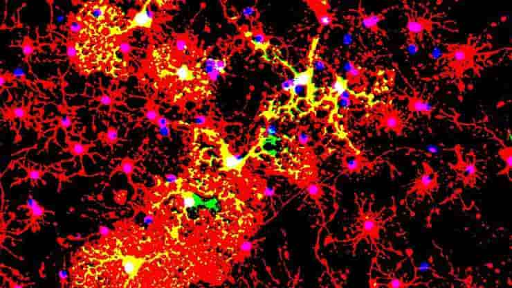
Image: Oligodendrocytes in culture. Credit: jakeyoung64 CC-BY
Last Updated on April 10, 2024