A surprisingly precise nerve cell connectivity pattern in the part of the cerebral cortex that is responsible for orienting the individual animal or human in space has been uncovered by a team of scientists around Moritz Helmstaedter at the Max Planck Institute for Brain Research.
Our brains house extremely complex neuronal circuits whose detailed structures are still largely unknown. This is especially true for the cerebral cortex of mammals, where, among other things, vision, thoughts or spatial orientation are computed.
Synapses in this region of the brain, the researchers report, are sorted very precisely along the electrical cables of the nerve cells. The nerve cells establish an unexpectedly precise circuit motive in which inhibitory nerve cells are contacted before the activation of the next nerve cell can be executed.
This nerve cell “trios” motif is a core connectivity motif in the cerebral cortex. Scientists speculate that such a highly precise circuit motive could be used for computing hypotheses about the next step in space.
Connectomics Of Grid Cells
Connectomics researchers have now used their repertoire of measuring and analysis techniques to study the medial entorhinal cortex, the part of the cerebral cortex in which grid cells provide a very particular representation of the space around the individual animal or human. These grid cells are active when the animal or human is located at highly ordered grid-like locations in a room or a large space.
Previously, scientists had already found a special arrangement of nerve cells in this region of the brain and had speculated that particular nerve cell circuits could exist within these special cell assemblies.
In the current study, the scientists looked at these circuits in more detail and found that, contrary to prior belief, the synapses are exceptionally precisely positioned. Within an extremely dense network of nerve cells, the nerve cells are, in fact, arranged in orderly triplets in which a nerve cell first activates an inhibitory nerve cell.
Transfer of the signal to the next excitatory nerve cell can, however, be hindered by the veto of the inhibitory nerve cell.
This core circuit, more or less functioning like a cortical transistor, would be able to propagate information in a very selective way, for instance, only when additional information about the context and the surroundings of the animal or the human is available.
The nerve cells within this transistor apparently use the very precise positioning of contact sites along their electrically conducting nerve cell cables, called axons.
“While many consider the cerebral cortex as a randomly assembled web of nerve cells and have already turned to simulating this random network, we now discover an extremely precise connectivity pattern. In the cerebral cortex, taking a much closer look is clearly worth it,”
says Helmstaedter.
Reference:
- Helene Schmidt, Anjali Gour, Jakob Straehle, Kevin M. Boergens, Michael Brecht & Moritz Helmstaedter. Axonal synapse sorting in medial entorhinal cortex. Nature (2017) doi:10.1038/nature24005
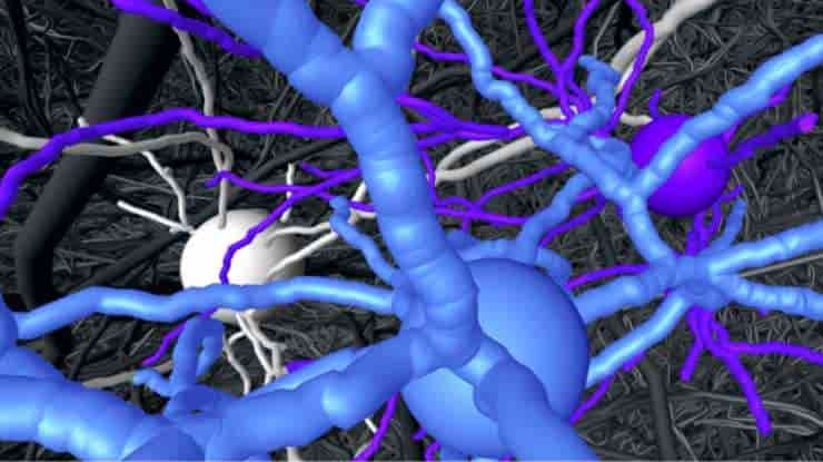
Image: Nerve cell “trio” (in color) found to be very specifically connected within the dense network of the brain (shown in grey). Credit: MPI for Brain Research
Last Updated on September 30, 2023